. 2013 Feb 5;110(6):2383-8.
doi: 10.1073/pnas.1215555110. Epub 2013 Jan 23.

UV light phototransduction activates transient receptor potential A1 ion channels in human melanocytes

Affiliations

UV light phototransduction activates transient receptor potential A1 ion channels in human melanocytes

Nicholas W Bellono et al. Proc Natl Acad Sci U S A. .

Abstract

Human skin is constantly exposed to solar ultraviolet radiation (UVR), the most prevalent environmental carcinogen. Humans have the unique ability among mammals to respond to UVR by increasing their skin pigmentation, a protective process driven by melanin synthesis in epidermal melanocytes. The molecular mechanisms used by melanocytes to detect and respond to long-wavelength UVR (UVA) are not well understood. We recently identified a UVA phototransduction pathway in melanocytes that is mediated by G protein-coupled receptors and leads to rapid calcium mobilization. Here we report that in human epidermal melanocytes physiological doses of UVR activate a retinal-dependent current mediated by transient receptor potential A1 (TRPA1) ion channels. The TRPA1 photocurrent is UVA-specific and requires G protein and phospholipase C signaling, thus contributing to UVA-induced calcium responses to mediate downstream cellular effects and providing evidence for TRPA1 function in mammalian phototransduction. Remarkably, TRPA1 activation is required for the UVR-induced and retinal-dependent early increase in cellular melanin. Our results show that TRPA1 is essential for a unique extraocular phototransduction pathway in human melanocytes that is activated by physiological doses of UVR and results in early melanin synthesis.


Conflict of interest statement

The authors declare no conflict of interest.

Figures

Fig. 1.
Fig. 1.
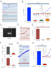
UVR activates a retinal- and dose-dependent UVA-specific current in HEMs. (A) UVR activates a retinal-dependent, outwardly rectifying current in HEMs that has a reversal potential near 0 mV. Whole-cell current amplitude at +80 mV in a representative HEM stimulated with 240 mJ/cm2 UVR (shaded box, 12 s of 20 mW/cm2) increased only when preincubated with all-trans retinal (12 μM). (B) The mean amplitude of the current density at +80 mV significantly increased when HEMs were incubated with 9-cis or all-trans retinal and stimulated with UVR (240 mJ/cm2), but not in the absence of retinal. IUVR = (5.29 ± 0.95) pA/pF for all-trans retinal and (6.54 ± 1.06) pA/pF for 9-cis retinal vs. (0.02 ± 0.09) pA/pF in the absence of retinal. n = 5–10 cells per condition, P < 0.0005, bars represent average ± SEM. (C) Current density was measured with a voltage step protocol in response to 240 mJ/cm2 UVR following incubation with 9-cis or all-trans retinal (12 μM); +80 mV was used for measuring the UVR-induced current under various conditions. (D) Representative whole-cell currents recorded at +80 mV from HEMs stimulated with increasing doses of UVR: I, 40 mJ/cm2 (20 mW/cm2 for 2 s); II, 140 mJ/cm2 (20 mW/cm2 for 7 s); III, 240 mJ/cm2 (20 mW/cm2 for 12 s); and IV, 340 mJ/cm2 (20 mW/cm2 for 17 s). The increase in current evoked by 340 mJ/cm2 UVR was blocked by RR (10 μM) (IV). The mean current density in response to 340 mJ/cm2 was significantly decreased in the presence of RR (10 μM). n = 4–5 cells per condition, ± SEM. (E) The amplitude of the UVR-induced (240 mJ/cm2) whole-cell current density in HEMs is dose-dependent. The current density of HEMs exposed to the UVR doses indicated in D were averaged for each dose, and the dose–response curve fit with a sigmoidal function. (F) The light-activated current in HEMs is UVA-specific. Mean amplitude of the increase in current density in HEMs stimulated with 250 mJ/cm2 UVR (∼90% UVA and ∼10% UVB), the UVB component (280–320 nm, 25 mJ/cm2), the UVA component (320–400 nm, 225 mJ/cm2), 250 mJ/cm2 blue (435–460 nm), or green (500–550 nm) light. UVA elicited a change in current density comparable to total UVR and much higher than UVB, blue or green. IUVR = (5.13 ± 0.48) pA/pF, IUVA = (4.85 ± 1.47) pA/pF, Iblue = (0.34 ± 0.28) pA/pF, Igreen = (0.19 ± 0.05) pA/pF. n = 5–6 cells per condition, P < 0.0001 for UVR vs. UVB, blue or green, ± SEM.
Fig. 2.
Fig. 2.
TRPA1 is expressed in HEMs and mediates the UVR-induced current. (A) Representative retinal-dependent HEM whole-cell currents at +80 mV in response to 240 mJ/cm2 UVR (20 mW/cm2 for 12 s) were reduced by TRPA1 antagonists RR (10 μM), camphor (1 mM), or HC-030031 (100 μM) compared with vehicle (0.5% DMSO). (B) The UVR-induced (240 mJ/cm2) increase in mean HEM whole-cell current density was significantly reduced by treatment with RR, camphor, or HC-030031 compared with vehicle. IUVR = (4.76 ± 0.91) pA/pF, IUVR/RR = (0.27 ± 0.25) pA/pF, IUVR/camphor = (0.36 ± 0.20) pA/pF, IUVR/HC-030031 = (0.79 ± 0.24) pA/pF. n = 6–8 cells per condition, P < 0.001, ± SEM. (Inset) UVR-induced increases in HEM current density as a function of voltage, in the presence of vehicle, RR, camphor, or HC-030031. (C) TRPA1 mRNA is expressed in HEMs. RT-PCR using HEM cDNA and TRPA1 specific primers identified a DNA band of the expected size that was analyzed and found to correspond to human TRPA1 cDNA. (D) Functional TRPA1 channels are present in HEMs. HEM whole-cell current measured at +80 mV was increased by TRPA1 agonist CA (500 μM), but not by coapplication of CA and TRPA1 antagonist HC-030031 (100 μM). (E) The mean whole-cell current density of HEMs was significantly increased by CA, but not when coapplied with RR (10 μM) or HC-030031 (100 μM). ICA = (8.46 ± 1.16) pA/pF, ICA/RR = (0.64 ± 0.08) pA/pF, ICA/HC-030031 = (0.08 ± 0.08) pA/pF. n = 4–5 cells per condition, P < 0.0007, ± SEM. (F) HEMs expressing TRPA1-targeted miRNA have reduced levels of TRPA1 mRNA and current. (Upper) Quantitative PCR (qPCR) analysis showed an ∼85% reduction in TRPA1 mRNA levels in HEMs expressing TRPA1-targeted miRNA, compared with control miRNA. n = 3 experiments, P < 0.002, ± SEM. (Lower) Mean CA-evoked (500 μM) current density at +80 mV in HEMs expressing TRPA1-targeted miRNA was ∼93% lower compared with control miRNA. ICA/ctrl miRNA = (6.56 ± 0.76) pA/pF, ICA/TRPA1 miRNA = (0.47 ± 0.22) pA/pF. n = 6 cells per condition, P < 0.0001, ± SEM. (G) Representative HEM expressing TRPA1-targeted miRNA exhibited a much smaller UVR-induced (240 mJ/cm2) increase in current at +80 mV than a HEM expressing control miRNA. (H) HEMs expressing TRPA1-targeted miRNA had a ∼92% lower peak UVR photocurrent density compared with those expressing control miRNA. IUVR/ctrl miRNA = (4.66 ± 0.86) pA/pF, IUVR/TRPA1 miRNA = (0.35 ± 0.12) pA/pF. n = 9–10 cells per condition, P < 0.0001, ± SEM (Inset) UVR-induced increase in current density vs. voltage in HEMs expressing control or TRPA1-targeted miRNA.
Fig. 3.
Fig. 3.
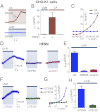
TRPA1 is activated downstream of a G protein and PLC-dependent signaling cascade. (A) TRPA1 expressed in CHO-K1 cells did not exhibit UVR- and retinal-dependent current. Whole-cell currents at +80 mV from representative CHO-K1 cells expressing TRPA1 were increased by 500 μM CA (Upper trace), but not by 240 mJ/cm2 UVR (Lower trace) following incubation with all-trans retinal. (B) The mean whole-cell current density in CHO-K1 cells increased significantly only in cells expressing TRPA1 and stimulated with CA, but not in cells expressing TRPA1, incubated with all-trans retinal, and stimulated with 240 mJ/cm2 UVR. ITRPA1/UVR+retinal = (0.46 ± 0.24) pA/pF, ITRPA1/CA = (28.48 ± 7.27) pA/pF, Iu/CA = (0.09 ± 0.17) pA/pF. n = 4–5 cells per condition, P < 0.008, ± SEM. (C) The CA-activated current in CHO-K1 cells expressing TRPA1 has a voltage-dependence similar to that of the UVR-induced current in HEMs (Fig. 1C). Stimulation of CHO-K1 cells expressing TRPA1 and incubated with all-trans retinal with 240 mJ/cm2 UVR did not evoke a current at any voltage. (D) The UVR-activated current is G protein-dependent. The UVR-induced (240 mJ/cm2) increase in currents at +80 mV in representative HEMs was abolished by intracellular GDPβS (1 mM) and by suramin (50 μM). (E) The UVR-induced (240 mJ/cm2) increase in mean HEM whole-cell current density was significantly reduced by dialyzing cells with 1 mM GDPβS or 50 μM suramin. IUVR = (4.80 ± 0.36) pA/pF, IUVR/GDPβS = (0.30 ± 0.20) pA/pF, IUVR/suramin = (0.58 ± 0.07) pA/pF. n = 6–7 cells per condition, P < 0.0001, ± SEM. (F) The UVR-activated current is PLC-dependent. The UVR-induced (240 mJ/cm2) increase in representative HEM whole-cell currents at +80 mV was abolished in the presence of the PLC inhibitor U73122 (9 μM), but not its inactive analog U73343 (9 μM). (G) UVR-induced increases in HEM current density as a function of voltage, in the presence of U73122 (9 μM) or its inactive analog U73343 (9 μM). (H) The UVR-induced (240 mJ/cm2) increase in mean HEM whole-cell current density was significantly reduced by treatment with U73122 (9 μM) compared with its inactive analog U73343 (9 μM). IUVR/U73122 = (6.21 ± 0.85) pA/pF, IUVR/U73343 = (0.86 ± 0.28) pA/pF. n = 5–6 cells per condition, P < 0.0002, ± SEM.
Fig. 4.
Fig. 4.
TRPA1 contributes to UVR-induced calcium responses. (A) Pseudochrome Fluo-4 fluorescence images of HEMs preincubated with all-trans retinal and stimulated with 240 mJ/cm2 UVR. (Upper) Images recorded in the presence of extracellular Ca2+ (+[Ca2+]ec), before UVR stimulation (−UVR) and at the peak UVR-induced Ca2+ response (+UVR). (Lower) Images recorded in the absence of extracellular Ca2+ (−[Ca2+]ec) and had a reduced response to UVR (Upper vs. Lower +UVR). (B) Representative Ca2+ imaging traces of HEMs preincubated with all-trans retinal and stimulated with UVR (240 mJ/cm2). The peak fluorescence intensity normalized to ionomycin (2 μM) (Fnorm) was higher in the presence of extracellular Ca2+ (+[Ca2+]ec) than in Ca2+-free solution (−[Ca2+]ec). Each trace is the average of 10–12 cells from one experiment. (C) In paired experiments, the normalized peak fluorescence of Ca2+ responses (Fnorm, max) of HEMs stimulated with 240 mJ/cm2 UVR in the presence of extracellular Ca2+ (+[Ca2+]ec) was higher than in the absence of extracellular Ca2+ (−[Ca2+]ec). Fnorm,max = (0.62 ± 0.04) for +[Ca2+]ec, Fnorm,max = (0.33 ± 0.05) for −[Ca2+]ec. n = 8 experiments per condition; each experiment is the average of 8–15 cells, P < 0.002. (D) The apparent free intracellular Ca2+ concentration ([Ca2+]ic) of representative UVR-evoked (240 mJ/cm2) retinal-dependent responses in the presence of extracellular Ca2+ (+[Ca2+]ec) was similarly reduced by HC-030031 (100 μM) and the absence of extracellular Ca2+ (−[Ca2+]ec). Incubation with thapsigargin (1 μM) in the absence of extracellular Ca2+ abolished UVR-induced Ca2+ responses. (E) The mean increase in apparent free [Ca2+]ic in response to 240 mJ/cm2 UVR in the presence of extracellular Ca2+ (+[Ca2+]ec) was reduced by ∼52% in the absence of extracellular Ca2+ (−[Ca2+]ec), by ∼57% in the presence 100 μM HC-030031, and by ∼93% following pretreatment with 1 μM thapsigargin in the absence of extracellular Ca2+. [Ca2+]ic,max = (994 ± 67) nM for +[Ca2+]ec, (479 ± 78) nM for −[Ca2+]ec, (429 ± 96) nM for HC-030031, (71 ± 21) nM for thapsigargin. n = 6–8 experiments per condition, P ≤ 0.003, ± SEM. (F) HEMs expressing TRPA1-targeted miRNA had a reduced retinal-dependent Ca2+ response to UVR (240 mJ/cm2), compared with HEMs expressing control miRNA, both stimulated in the presence of extracellular Ca2+. n = 4–8 cells per condition. (G) The mean peak increase in apparent free [Ca2+]ic (nM) induced by 240 mJ/cm2 UVR in HEMs expressing TRPA1-targeted miRNA was reduced by ∼58% compared with control miRNA. [Ca2+]ic,max = (737 ± 117) nM for control, (307 ± 70) nM for TRPA1-targeted miRNA. n = 5 experiments per condition, P < 0.001, ± SEM.
Fig. 5.
Fig. 5.
TRPA1 is required for UVR-induced early melanin synthesis. (A) The UVR-induced retinal-dependent increase in melanin was reduced by 78.7 ± 12.5% in HEMs exposed to 2.5 J/cm2 UVR in the presence of 100 μM HC-030031 compared with vehicle control (0.5% DMSO). Relative melanin increase measured 8 h after UVR exposure of HEMs preincubated with all-trans retinal, and calculated relative to melanin increase in cells receiving identical treatment without exposure to UVR. n = 12 experiments, P < 0.0001, ± SEM. (B) The retinal-dependent UVR-induced peak Ca2+ response in HEMs stimulated with 2.5 J/cm2 UVR (20 mW/cm2 for 125 s) in the presence of HC-030031 (100 μM) was reduced by ∼36.7%, but the sustained elevation measured 275 s after UVR stimulation was reduced by ∼70%. n ≥ 25 cells per condition, P < 0.007 for differences in peak Ca2+ response and P < 0.02 for differences in sustained Ca2+ elevation for n = 6 experiments. (C) The retinal-dependent increase in melanin in response to 2.5 J/cm2 UVR measured 8 h after exposure was abolished in HEMs expressing TRPA1-targeted miRNA compared with control miRNA. The melanin increase was calculated relative to changes in melanin measured in nonirradiated cells preincubated with retinal and expressing the corresponding miRNA. n = 6 experiments per condition, P < 0.0007, ± SEM.

References

    1. Lin JY, Fisher DE. Melanocyte biology and skin pigmentation. Nature. 2007;445(7130):843–850. - PubMed
    1. Kobayashi N, et al. Supranuclear melanin caps reduce ultraviolet induced DNA photoproducts in human epidermis. J Invest Dermatol. 1998;110(5):806–810. - PubMed
    1. Cosens DJ, Manning A. Abnormal electroretinogram from a Drosophila mutant. Nature. 1969;224(5216):285–287. - PubMed
    1. Montell C, Jones K, Hafen E, Rubin G. Rescue of the Drosophila phototransduction mutation trp by germline transformation. Science. 1985;230(4729):1040–1043. - PubMed
    1. Hardie RC, Minke B. The trp gene is essential for a light-activated Ca2+ channel in Drosophila photoreceptors. Neuron. 1992;8(4):643–651. - PubMed

Publication types

MeSH terms