. 2013 Feb 19;110(8):E668-77.
doi: 10.1073/pnas.1221484110. Epub 2013 Jan 23.

Arl1p regulates spatial membrane organization at the trans-Golgi network through interaction with Arf-GEF Gea2p and flippase Drs2p

Affiliations

Arl1p regulates spatial membrane organization at the trans-Golgi network through interaction with Arf-GEF Gea2p and flippase Drs2p

Pei-Chin Tsai et al. Proc Natl Acad Sci U S A. .

Abstract

ADP ribosylation factors (Arfs) are the central regulators of vesicle trafficking from the Golgi complex. Activated Arfs facilitate vesicle formation through stimulating coat assembly, activating lipid-modifying enzymes and recruiting tethers and other effectors. Lipid translocases (flippases) have been implicated in vesicle formation through the generation of membrane curvature. Although there is no evidence that Arfs directly regulate flippase activity, an Arf-guanine-nucleotide-exchange factor (GEF) Gea2p has been shown to bind to and stimulate the activity of the flippase Drs2p. Here, we provide evidence for the interaction and activation of Drs2p by Arf-like protein Arl1p in yeast. We observed that Arl1p, Drs2p and Gea2p form a complex through direct interaction with each other, and each interaction is necessary for the stability of the complex and is indispensable for flippase activity. Furthermore, we show that this Arl1p-Drs2p-Gea2p complex is specifically required for recruiting golgin Imh1p to the Golgi. Our results demonstrate that activated Arl1p can promote the spatial modulation of membrane organization at the trans-Golgi network through interacting with the effectors Gea2p and Drs2p.


Conflict of interest statement

The authors declare no conflict of interest.

Figures
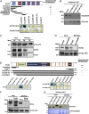
Fig. 1.
Fig. 1.
Arl1p interacts with Gea2p and Drs2p. (A) The N-terminal region of Gea2p interacts with active Arl1p in a yeast two-hybrid assay. (Upper) Schematic diagram of Gea2 truncation mutant constructs. (Lower) pEG202 plasmids containing Arl1dN17Q72L or Arl1dN17T32N were cotransformed with pJG4-5 plasmids containing different forms of Gea2p into yeast, and their interactions were analyzed with reporter β-galactosidase activity. (B) Gea2p and Arl1p specifically and directly interact with each other in vitro. Purified recombinant His-Gea2N250 was incubated with GST-Arfs as indicated for 1 h at 4 °C. After the wash, the bound Gea2 proteins were detected with anti-His antibody. (C) Active Arl1p forms a complex with Gea2p and Drs2p in vivo. Constitutively active or inactive Arl1-GFP was immunoprecipitated from yeast, which ectopically expressed Drs2p and 3HA-Gea2p. Bound Gea2p and Drs2p were detected with Western blotting. (D) The N-terminal region of Gea2p is required for interaction with Arl1p. Gea2p or Gea2pdN89 was coexpressed with Arl1-GFP in arl1-null cells. Arl1-GFP was immunoprecipitated, and the bound Gea2p was detected with anti-Gea2p antibody. (E) Interactions between Arf proteins and Drs2p. (Upper) Schematic diagram of Drs2-truncation mutant constructs. Orange bar, transmembrane domain. (Lower) pEG202 encoding various Arf proteins and pJG4-5 encoding Drs2p were cotransformed into yeast, and their interactions were determined using a β-galactosidase assay. (F) The N-terminal region of Drs2p is required for interaction with the active form of Arl1p. Drs2 or Drs2dN160 was transformed into arl1-null cells expressing Arl1QL-GFP. The Arl1-GFP was precipitated with anti-GFP antibody, and the bound Drs2p was detected with anti-Drs2p antibody. (G) Drs2p and Arl1p specifically and directly interact with each other in vitro. One microgram of purified GST-Drs2N220 was incubated with 1 μg of His-Arl proteins. After the wash, bound small G proteins were detected with anti-His antibody. Asterisk indicates the degraded form of Drs2N220.
Fig. 2.
Fig. 2.
Arl1p, Drs2p, or Gea2p is required for interaction between the other proteins. (A) Arl1p affects the interaction between Gea2p and Drs2p. Drs2p was ectopically expressed in drs2Δ or drs2Δarl1Δ cells, which contained chromosomally HA-tagged Gea2p under the GAL1 promoter. HA-Gea2 was immunoprecipitated by anti-HA antibody, and the coprecipitated Drs2p was detected with anti-Drs2p antibody. The interaction efficiency of Drs2p and 3HA-Gea2p was normalized to 3HA-Gea2p. The quantitative result is shown (Right). (B) Drs2p is required for Arl1p to interact with Gea2p. Ha-Gea2p was coexpressed with constitutively active or inactive Arl1p in WT or drs2Δ cells. HA-Gea2p was immunoprecipitated with anti-HA antibody, and bound Arl1p was detected with immunoblotting. The relative interaction of Arl1p and HA-Gea2p was quantified and normalized to the amount of HA-Gea2p (Right). (C) The interaction of Arl1p with Drs2p requires Gea2p. Drs2p and constitutively active or inactive Arl1-GFP were ectopically expressed in WT or gea2Δ cells. Arl1-GFP was immunoprecipitaed with anti-GFP antibody, and the bound Drs2p was detected with immunoblotting. The bound Drs2p was normalized to Arl1-GFP and is shown (Right). These results (A–C) are presented as the mean ± SD of three independent experiments (*P < 0.005; t test).
Fig. 3.
Fig. 3.
Drs2p and Gea2p are involved in the localization of Imh1p and Gas1p. (A) Gea2p and Drs2p are required for the proper distribution of Imh1p to the TGN membrane. mCherry-Imh1 and GFP-Sft2 were cotransformed into different mutant yeast as indicated. Live cell images were acquired from midlog phase cultured yeast by fluorescence microscopy. (B) Gea2p and Drs2p affect the transport of Gas1p. Gas1-GFP was expressed in different mutant yeast strains; live cells in the midlog phase were observed by fluorescence microscopy. (C) Loss of GEA2 or DRS2 results in phenotypes that are similar to arl1Δ cells. WT and mutant yeast strains were grown to the stationary phase, and serial dilutions were spotted onto plates. (D) Arl1p, but not Drs2p or Gea2p, is required for the subcellular distribution of GFP-Gga2. GFP-Gga2 was transformed into WT, arl1Δ, gea2Δ, drs2Δ, or mon2Δ yeast. Yeast cultures in the midlog phase were observed by fluorescence microscopy. (Scale bar: 5 μm.)
Fig. 4.
Fig. 4.
The interaction between Arl1p and Gea2p or Drs2p is required for the Golgi localization of Imh1p. (A) Effect of Drs2dN160 on Imh1p localization. Drs2p or various Drs2p mutants were individually coexpressed with Imh1-mCherry in drs2Δ cells. The Imh1-mCherry distribution was observed in the indicated midlog phase yeasts by fluorescence microscopy. (Scale bar: 5 μm.) (B) The interaction between Arl1p and Drs2p is not required for the transport of Gas1p. Gas1-GFP was coexpressed with full-length Drs2p or Drs2 mutants in drs2Δ, and live cells in the midlog phase were observed by fluorescence microscopy. (Scale bar: 5 μm.) (C) The ability of Drs2p truncation to rescue cold and Congo Red hypersensitivity in drs2Δ. drs2-null cells expressing different truncated Drs2p were serially diluted, spotted onto plates, and cultured as indicated. (D) The N-terminal region of Gea2p is required for the localization of Imh1p. Localization of mCherry-Imh1 in gea2-null yeast expressing different forms of Gea2p is shown. (Scale bar: 5 μm.) (E) The interaction between Arl1p and Gea2p is dispensable for the transport of Gas1p. The localization of Gas1-GFP in gea2Δ yeast expressing different forms of Gea2p was observed by fluorescence microscopy. (Scale bar: 5 μm.) (F) Complementation of gea2-null cells with plasmids carrying GEA2, GEA2dN89, or GEA2dN250. Cells were serially diluted and spotted onto plates with or without Congo Red. (G) Gea2dN89 rescues the temperature sensitivity of gea2gea1ts cells. Different forms of GEA2 were transformed into gea2gea1-6ts yeast. The cells were serially diluted, spotted onto plates, and incubated at 25 °C or 37 °C.
Fig. 5.
Fig. 5.
The flippase activity of Golgi membranes requires Arl1p. (A) The activity of Drs2p is required for the localization of Imh1p. The localization of mCherry-Imh1 in drs2Δ expressing different forms of Drs2p is shown. (Scale bar: 5 μm.) (B–E) The flippase activity of the Golgi membrane from different yeasts. Golgi membranes from WT (B), arl1Δ (C), gea2Δ (D), and drs2Δ (E) were assayed for NBD-PS flippase activity at 30 °C, as described in Materials and Methods. The arrow indicates the addition of ATP or the mock control, and the difference in the NBD-PS in the cytosolic leaflet between the mock or ATP addition was considered to be 100% NBD-PS flippase activity (asterisk). (F) Quantification of NBD-PS flippase activity. The flippase activity for NBD-PS on the Golgi membranes from the indicated yeast strains was normalized to WT membranes, which was set at 100%. (G) Arl1p directly regulates the flippase activity of the Golgi membrane. The NBD-PS flippase activity of Golgi membrane from arl1Δ was measured with or without the addition of purified recombinant Arl1, Arl1Q72L, or Arl1T32N. (H) The N-terminal region of Drs2p is required for the flippase activity of Drs2p. The NBD-PS flippase activity of the Golgi membranes purified from WT cells and drs2Δ cells expressing Drs2p or Drs2dN160. (I) The N-terminal region of Gea2p is important for the translocation of NBD-PS at the TGN. Shown is the NBD-PS flippase activity in TGN membranes purified from WT cells and gea2Δ cells expressing Gea2p or Gea2dN89. All of the data (B–I) represent the mean ± SD (n = 3; *P < 0.001; t test).
Fig. 6.
Fig. 6.
Gea2p, Arl1p, and Drs2p form a protein complex that affects the membrane dynamics and lipid asymmetry of the Golgi. The binding of Arl1p to the N-terminal and Gea2p to the C-terminal regions of Drs2p stimulate the ATP-dependent flippase activity of Drs2p at the TGN membrane. Active Drs2p increases the surface area of the cytosolic leaflet at the expense of the luminal leaflet and bends the membrane toward the cytosol. Then, Imh1p is able to interact with the membrane and retains its Golgi localization. Moreover, the deletion of GEA2 or DRS2 impairs Arl1-Drs2-Gea2 protein complex formation, which affects the PS asymmetry of the TGN and the Golgi localization of Imh1p.

Comment in

Similar articles

Cited by

References

    1. D’Souza-Schorey C, Chavrier P. ARF proteins: Roles in membrane traffic and beyond. Nat Rev Mol Cell Biol. 2006;7(5):347–358. - PubMed
    1. Gillingham AK, Munro S. The small G proteins of the Arf family and their regulators. Annu Rev Cell Dev Biol. 2007;23:579–611. - PubMed
    1. Kahn RA, et al. Nomenclature for the human Arf family of GTP-binding proteins: ARF, ARL, and SAR proteins. J Cell Biol. 2006;172(5):645–650. - PMC - PubMed
    1. Donaldson JG, Jackson CL. ARF family G proteins and their regulators: Roles in membrane transport, development and disease. Nat Rev Mol Cell Biol. 2011;12(6):362–375. - PMC - PubMed
    1. Pasqualato S, Renault L, Cherfils J. Arf, Arl, Arp and Sar proteins: A family of GTP-binding proteins with a structural device for ‘front-back’ communication. EMBO Rep. 2002;3(11):1035–1041. - PMC - PubMed

Publication types

MeSH terms

Substances