

doi: 10.1128/JVI.03235-12.
Epub 2013 Jan 23.
Susceptibility of human iris stromal cells to herpes simplex virus 1 entry
Affiliations
- PMID: 23345512
- PMCID: PMC3624200
- DOI: 10.1128/JVI.03235-12
Item in Clipboard
Susceptibility of human iris stromal cells to herpes simplex virus 1 entry
J Virol.
2013 Apr.
Abstract
Ocular herpes simplex virus 1 (HSV-1) infection can lead to multiple complications, including iritis, an inflammation of the iris. Here, we use human iris stroma cells as a novel in vitro model to demonstrate HSV-1 entry and the inflammatory mediators that can damage the iris. The upregulated cytokines observed in this study provide a new understanding of the intrinsic immune mechanisms that can contribute to the onset of iritis.
Figures
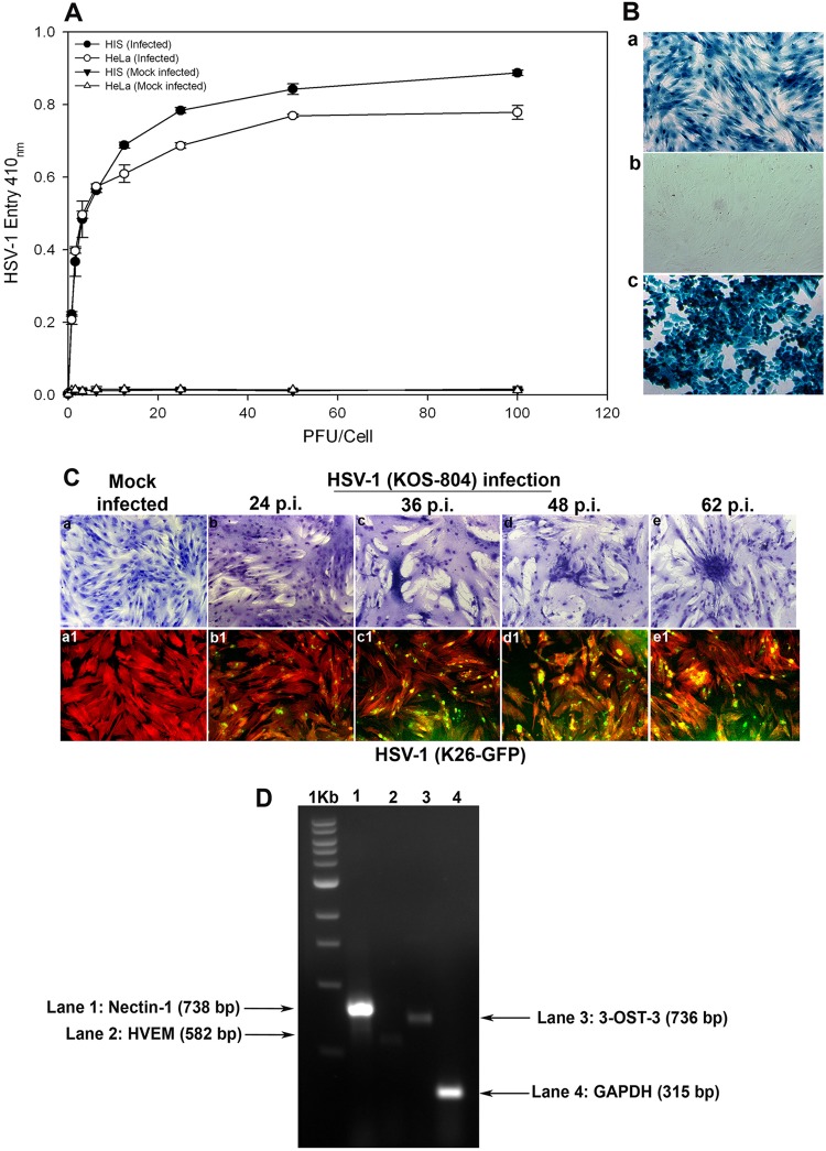
Analysis of herpes simplex virus 1 (HSV-1) entry in primary cultures of HIS cells. (A) Entry of HSV-1 into cultured HIS cells. Cultured HIS cells, along with HeLa cells, were plated in 96-well plates and inoculated with serial dilutions of a recombinant form of HSV-1 (KOS) gL86 that expresses β-galactosidase following cell entry at the indicated PFU/cell. After 6 h, the cells were washed, permeabilized, and incubated with o-nitrophenyl-β-d -galactopyranoside (ImmunoPure ONPG; Pierce) substrate for quantitation of β-galactosidase activity expressed from the input viral genome. The enzymatic activity was measured by spectrophotometer (Molecular Devices) at an optical density (OD) at 410 nm. Values in the figure were plotted as the means from three determinations (±standard deviations [SD]). (B) X-Gal staining of HSV-1 entry. Cultured HIS cells inoculated with HSV-1 (KOS) gL86 at an MOI of 0.01 (a) were 100% stained blue, indicating 100% viral infection, whereas uninfected cells (b) did not demonstrate any staining. (c) HeLa cells, with HSV-1 KOS gL86-infected cells stained blue, were used as a positive control. (C) Viral cell-to-cell spread. HIS cells were inoculated with serial dilutions of two recombinant forms of HSV-1, (KOS) 804, a mutant known to form high numbers of syncytia, and green fluorescent protein-tagged HSV-1 (K26) GFP. Confluent monolayer cultures inoculated with HSV-1 (KOS) 804 for 2 h were then washed and incubated with methyl cellulose containing media and stained at the various time points p.i. as indicated (a to e). Cells inoculated with HSV-1 (K26) GFP were subjected to fluorescence imaging (a1 to e1). Confluent monolayer cultures were stained with TRITC-conjugated phalloidin (red color), which binds to F-actin. HSV-1-infected cells showed both green fluorescence from HSV-1 capsid and red fluorescence from F-actin expression (green and red overlapping areas appear yellow). The images were taken with a fluorescence microscope at 20× magnification. (D) Standard nonquantitative RT-PCR analysis for entry receptor expression. Total RNA samples were isolated from HIS cells and converted to first-strand cDNAs and analyzed by PCR for the receptors as indicated. A housekeeping gene, GAPDH, was used as a control.
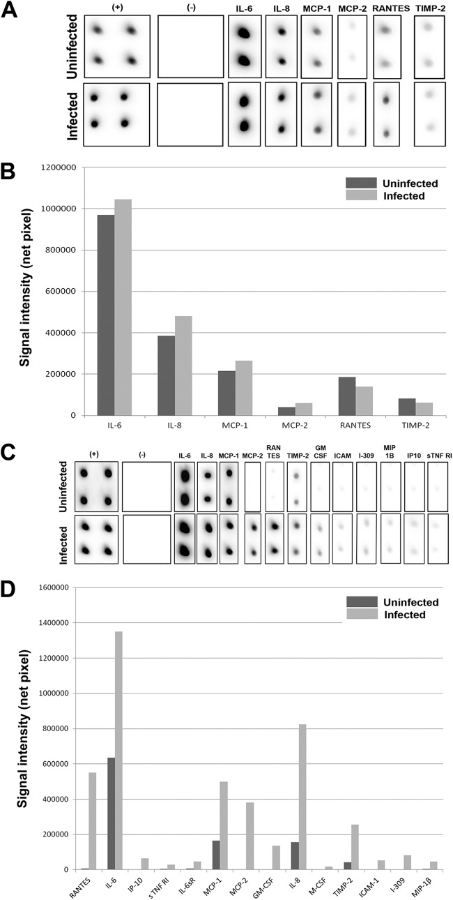
Inflammation in HIS cells upon HSV-1 infection. (A) A RayBio cytokine antibody array (RayBio, Norcross, GA) per the manufacturer's instructions was used to assess inflammatory markers produced in HSV-1 (KOS) 804 (MOI of 0.01) infection of an in vitro HIS cell culture at 1 day p.i.; (B) signal intensities of various inflammatory markers are plotted in uninfected versus infected cells at 1 day p.i.; (C) inflammatory markers measured at 2 days p.i. utilizing same material and methods as described above; (D) signal intensities of various inflammatory markers are plotted in uninfected versus infected cells at 2 days p.i.
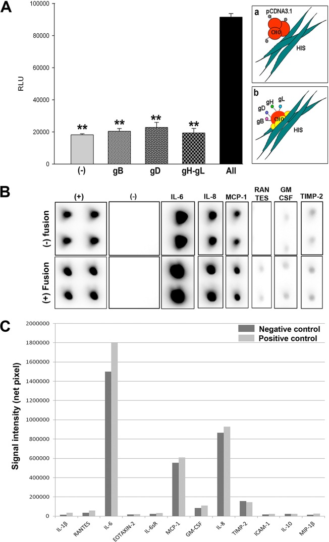
Cytokine response following membrane fusion. (A) HIS cell fusion with HSV-1 glycoprotein-expressing Chinese hamster ovary (CHO-K1) cells. CHO-K1 cells were either transfected with an empty vector (pCDNA3.1) or plasmids expressing one or more of the glycoproteins gB, gD, or gH-gL. A luciferase-based reporter system was used to measure fusion. Relative luciferase activity was measured in relative luciferase units (RLU) (y axis). Cell fusion was measured in RLU. CHO-K1 cells expressing the empty vector were used as a negative control. The schematic shows both unsuccessful (a) and successful (b) cell fusion between CHO-K1 and HIS cells. (B) A RayBio cytokine antibody array (RayBio, Norcross, GA) per the manufacturer's instructions was used to assess inflammatory markers produced in HIS cell interactions with CHO-K1 cells in test versus negative-control cases. (C) Signal intensities of various inflammatory markers are plotted in negative-control (no fusion) versus test (fusion) cells.
Similar articles
-
A synthetic glycosaminoglycan mimetic blocks HSV-1 infection in human iris stromal cells.Antiviral Res. 2019 Jan;161:154-162. doi: 10.1016/j.antiviral.2018.11.007. Epub 2018 Nov 24. Antiviral Res. 2019. PMID: 30481525
-
Herpes simplex virus type-1 (HSV-1) entry into human mesenchymal stem cells is heavily dependent on heparan sulfate.J Biomed Biotechnol. 2011;2011:264350. doi: 10.1155/2011/264350. Epub 2011 Jun 21. J Biomed Biotechnol. 2011. PMID: 21799659 Free PMC article.
-
Non-muscle myosin IIA is a functional entry receptor for herpes simplex virus-1.Nature. 2010 Oct 14;467(7317):859-62. doi: 10.1038/nature09420. Nature. 2010. PMID: 20944748
-
Anti-heparan sulfate peptides that block herpes simplex virus infection in vivo.J Biol Chem. 2011 Jul 15;286(28):25406-15. doi: 10.1074/jbc.M110.201103. Epub 2011 May 19. J Biol Chem. 2011. PMID: 21596749 Free PMC article.
-
Herpesvirus entry mediator on radiation-resistant cell lineages promotes ocular herpes simplex virus 1 pathogenesis in an entry-independent manner.mBio. 2015 Oct 20;6(5):e01532-15. doi: 10.1128/mBio.01532-15. mBio. 2015. PMID: 26489863 Free PMC article.
Cited by
-
HSV-1 interaction to 3-O-sulfated heparan sulfate in mouse-derived DRG explant and profiles of inflammatory markers during virus infection.J Neurovirol. 2017 Jun;23(3):483-491. doi: 10.1007/s13365-017-0521-4. Epub 2017 Mar 21. J Neurovirol. 2017. PMID: 28326469 Free PMC article.
-
Iridian anterior segment OCT in rubella uveitis syndrome and cytomegalovirus anterior uveitis: a comparative study.Graefes Arch Clin Exp Ophthalmol. 2022 Nov;260(11):3647-3655. doi: 10.1007/s00417-022-05733-3. Epub 2022 Jun 16. Graefes Arch Clin Exp Ophthalmol. 2022. PMID: 35708847
-
A role for 3-O-sulfated heparan sulfate in promoting human cytomegalovirus infection in human iris cells.J Virol. 2015 May;89(9):5185-92. doi: 10.1128/JVI.00109-15. Epub 2015 Feb 25. J Virol. 2015. PMID: 25717110 Free PMC article.
-
Induction of Filopodia During Cytomegalovirus Entry Into Human Iris Stromal Cells.Front Microbiol. 2022 Apr 5;13:834927. doi: 10.3389/fmicb.2022.834927. eCollection 2022. Front Microbiol. 2022. PMID: 35450284 Free PMC article.
-
High Preventive Effect of G2-S16 Anionic Carbosilane Dendrimer against Sexually Transmitted HSV-2 Infection.Molecules. 2020 Jun 28;25(13):2965. doi: 10.3390/molecules25132965. Molecules. 2020. PMID: 32605185 Free PMC article. Review.
References
-
- Dawson CR, Togni B. 1976. Herpes simplex eye infections: clinical manifestations, pathogenesis and management. Surv. Ophthalmol. 21:121–135 - PubMed
-
- Rathinam SR, Namperumalsamy P. 2007. Global variation and pattern changes in epidemiology of uveitis. Indian J. Ophthalmol. 55:173–183 - PubMed
-
- Teitelbaum CS, Streeten BW, Dawson CR. 1987. Histopathology of herpes simplex virus keratouveitis. Curr. Eye Res. 6:189–194 - PubMed
-
- Sungur GK, Hazirolan D, Yalvac IS, Ozer PA, Aslan BS, Duman S. 2010. Incidence and prognosis of ocular hypertension secondary to viral uveitis. Int. Ophthalmol. 30:191–194 - PubMed
-
- Tugal-Tutkun I, Ötük-Yasar B, Altinkurt E. 2010. Clinical features and prognosis of herpetic anterior uveitis: a retrospective study of 111 cases. Int. Ophthalmol. 30:559–565 - PubMed
Publication types
MeSH terms
Substances
Grants and funding
LinkOut - more resources
Full Text Sources
Other Literature Sources
