. 2013 Jan 23;5(169):169ra12.
doi: 10.1126/scitranslmed.3004688.

KLF6-SV1 drives breast cancer metastasis and is associated with poor survival

Affiliations

KLF6-SV1 drives breast cancer metastasis and is associated with poor survival

Raheleh Hatami et al. Sci Transl Med. .

Abstract

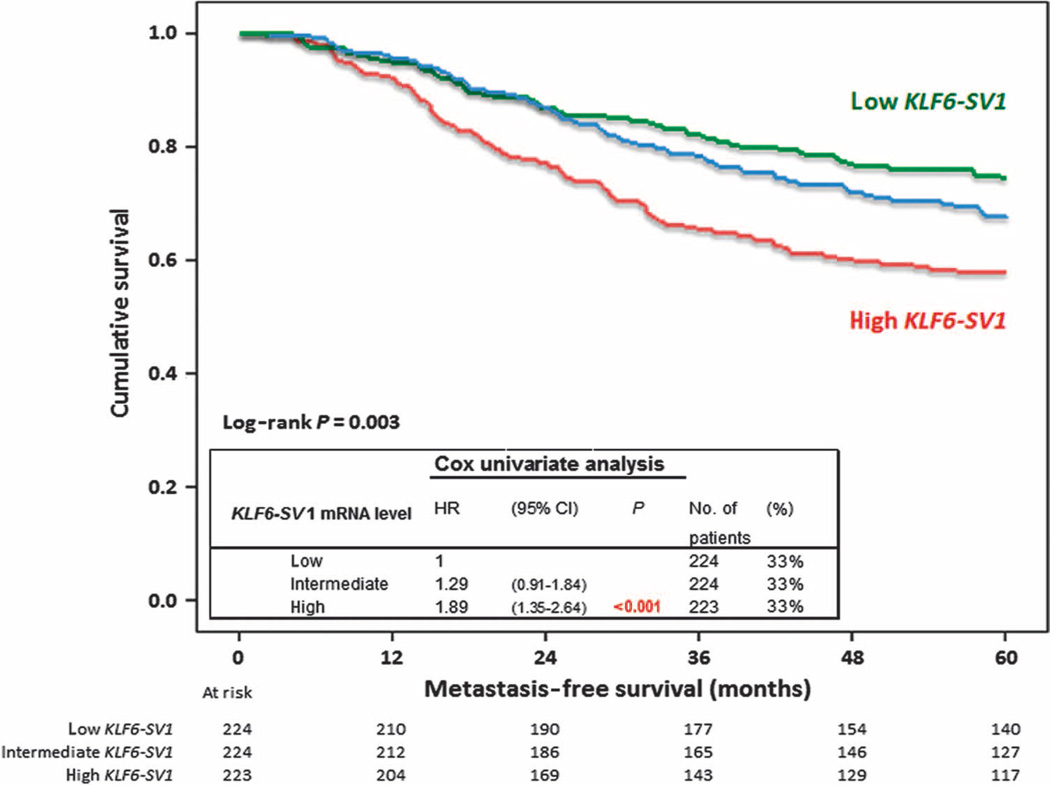
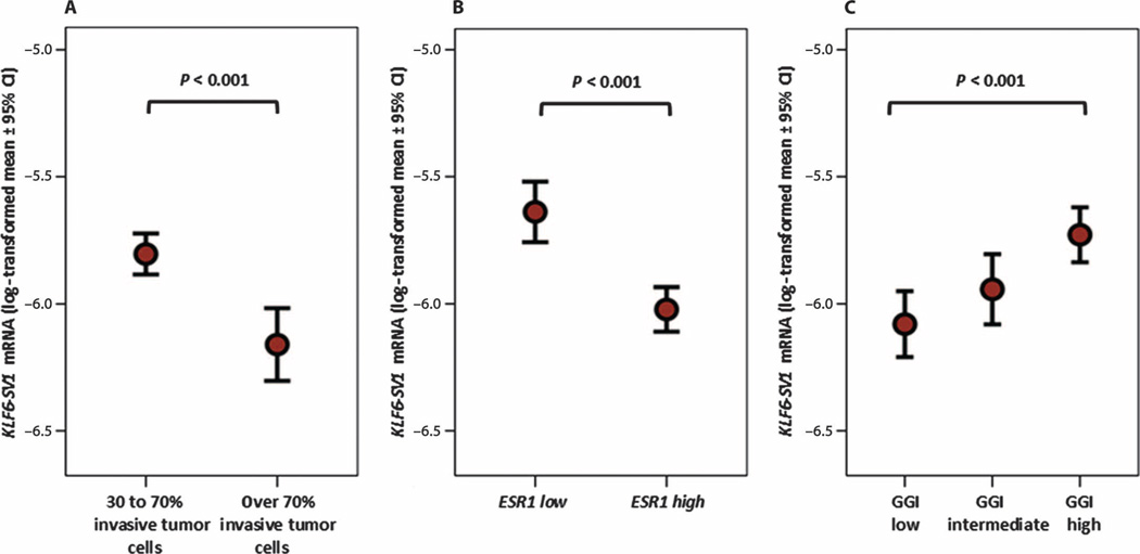
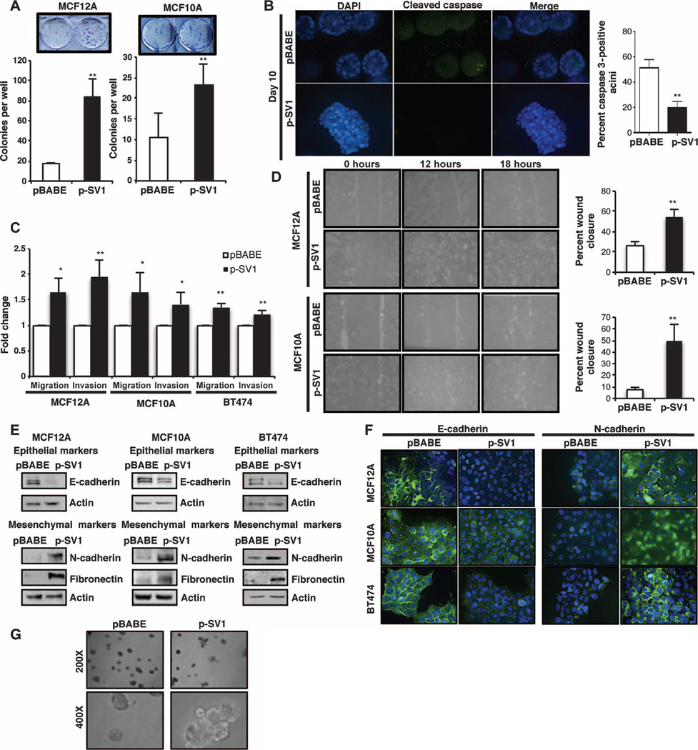
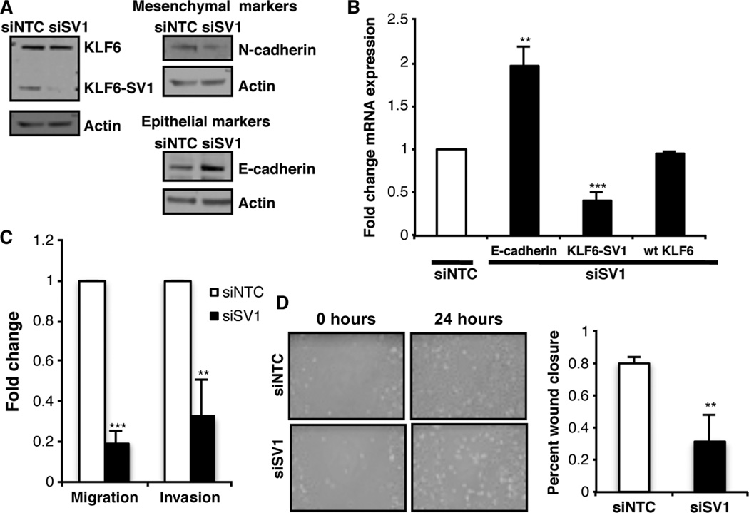
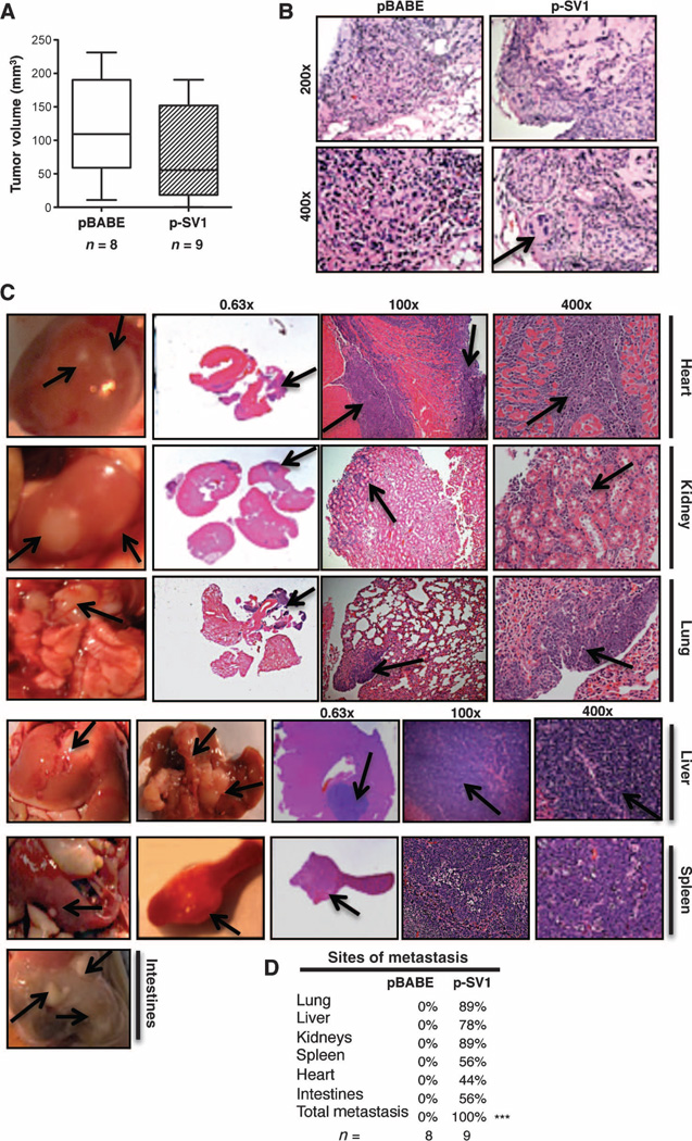
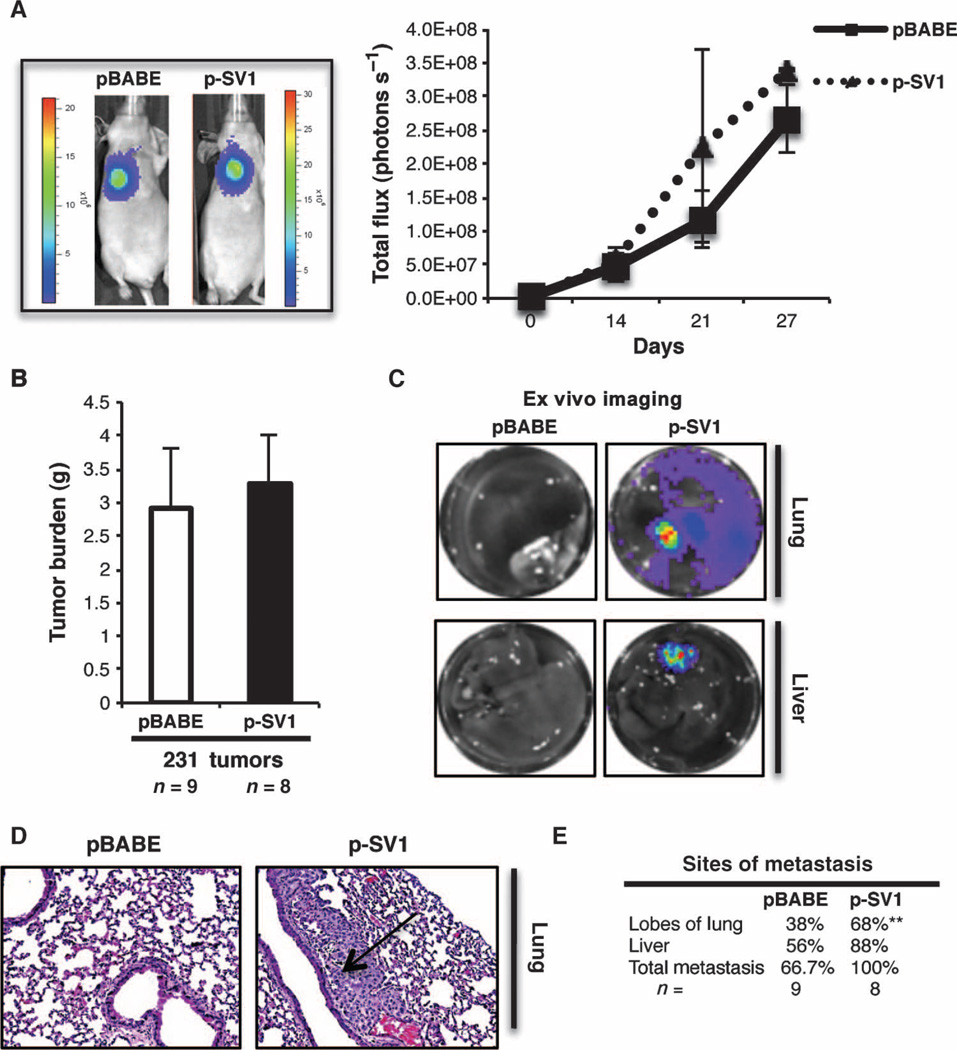
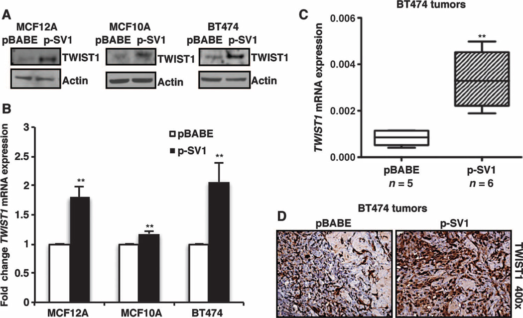
Metastasis is the major cause of cancer mortality. A more thorough understanding of the mechanisms driving this complex multistep process will aid in the identification and characterization of therapeutically targetable genetic drivers of disease progression. We demonstrate that KLF6-SV1, an oncogenic splice variant of the KLF6 tumor suppressor gene, is associated with increased metastatic potential and poor survival in a cohort of 671 lymph node-negative breast cancer patients. KLF6-SV1 overexpression in mammary epithelial cell lines resulted in an epithelial-to-mesenchymal-like transition and drove aggressive multiorgan metastatic disease in multiple in vivo models. Additionally, KLF6-SV1 loss-of-function studies demonstrated reversion to an epithelial and less invasive phenotype. Combined, these findings implicate KLF6-SV1 as a key driver of breast cancer metastasis that distinguishes between indolent and lethal early-stage disease and provides a potential therapeutic target for invasive breast cancer.


Figures

Fig. 1
Fig. 1
Increased KLF6-SV1SV1 expression in primary breast tumors correlates with poor prognosis. Five-year MFS as a function of KLF6-SV1 in 671 LNN primary breast cancer patients after trichotomizing KLF6-SV1 mRNA levels in three equal parts (low, intermediate, and high). Patients at risk are indicated.
Fig. 2
Fig. 2
High levels of KLF6-SV1 mRNA associated with primary tumor characteristics. (A) KLF6-SV1 mRNA levels were correlated with stromal content (n = 453 LNN primary breast tumors with 30 to 70% invasive tumor cells versus n = 218 with more than 70% invasive tumor cells). (B) ESR1 (n = 492 ESR1-positive LNN primary breast tumors versus n = 179 ESR1-negative LNN primary breast tumors). (C) GGI (n = 218 GGI low LNN primary breast tumors versus n = 220 GGI intermediate LNN primary tumors versus n = 221 GGI high LNN primary breast tumors). mRNA levels were measured by qRT-PCR. The strengths of the associations between continuous variables were tested with a two-tailed Mann-Whitney U test.
Fig. 3
Fig. 3
Overexpression of KLF6-SV1 promotes breast cancer development and progression in breast cell lines. (A) Colony assay of KLF6-SV1–expressing mammary epithelial cells stained at day 8; graphs represent the average number of colonies in pBABE control versus KLF6-SV1–expressing cells for MCF12A (P = 0.0027) and MCF10A (P = 0.0066). (B) Quantification of apoptosis in KLF6-SV1–expressing acini detected by cleaved caspase 3 staining by day 10 using indirect immunofluorescence (images taken at ×400 magnification). Acini containing two or more active caspase 3–positive cells were scored as positive, and 100 acini were counted per experiment (P = 0.0026). (C) Quantification of invasion and migration in Transwell chambers toward chemoattractant serum media over 24 hours in pBABE control versus KLF6-SV1–expressing cells in MCF12A migration (P = 0.034) and invasion (P = 0.002), MCF10A migration (P = 0.045) and invasion (P = 0.037), and BT474 migration (P = 0.011) and invasion (P = 0.0017). (D) Wound-healing assay over a period of 18 hours; graph represents percent wound closure over 12 hours for MCF12A (P = 0.006) and MCF10A (P = 0.007); images were taken at ×200 magnification. (E) Expression of epithelial marker E-cadherin and mesenchymal marker N-cadherin and fibronectin-expressing cells detected by Western blot. (F) Immunocytochemistry for N-cadherin and E-cadherin in pBABE versus p-SV1 cells taken at ×400 magnification. (G) KLF6-SV1–expressing MCF10A multiacinar structures during morphogenesis at day 20, taken at ×200 and ×400 magnifications. Representative images are demonstrated here and were repeated in triplicate; 1:100 of each antibody was used for staining. Data are represented as means ± SD of triplicate experiments.
Fig. 4
Fig. 4
Silencing KLF6-SV1 in metastatic cell line reverts cells back to an epithelial-like expression pattern. (A) Western blot analysis of wtKLF6, KLF6-SV1, N-cadherin, and E-cadherin expression after siRNA-mediated knockdown of KLF6-SV1 in MDA-MB-231-luc-D3H2LN cell line. (B) Gene expression analysis of KLF6-SV1 (P < 0.001) and E-cadherin (P = 0.002) analyzed by qRT-PCR. (C) Invasion (P < 0.001) and migration (P = 0.003) toward serum media over 24 hours. (D) Wound-healing assay from cells grown on a monolayer over 24 hours; graph represents percent wound closure over a period of 24 hours; images were taken at ×200 magnification (P = 0.007). Data are represented as means ± SD of triplicate experiments.
Fig. 5
Fig. 5
KLF6-SV1 induces metastasis in a subcutaneous mouse model. (A) Tumor volume of BT474 pBABE-expressing (n = 8) compared to p-SV1–expressing (n = 9) tumors. (B) Representative H&E staining of pBABE and p-SV1 tumors, with arrow pointing to local invasion in the p-SV1 tumor. (C) Representative images and H&E-stained sections of cardiac myocytes of the heart, kidney, lung, liver, spleen, and intestinal tract from a BT474 KLF6-SV1 mouse. Arrows point to metastatic lesions. (D) Sites of metastasis in BT474 pBABE- versus p-SV1–injected mice in the KLF6-SV1 mice (P < 0.001) analyzed by χ2 analysis. All data are represented as means ± SD.
Fig. 6
Fig. 6
Overexpression of KLF6-SV1 in an orthotopic model of breast cancer progression enhances metastasis. (A) Female nu/nu mice injected orthotopically with MDA-MB-231-luc-D3H2LN pBABE (n = 9) or p-SV1 (n = 8) cells into mammary fat pad; representative luminescent images from each group at day 21 and bioluminescence graph of local tumor growth for 27 days. (B) Tumor burden of pBABE (n = 9) and p-SV1 (n = 8) mammary tumors from mice sacrificed at day 27. (C) Representative ex vivo imaging of lung and liver from each group. (D) Representative H&E-stained lung section, with arrow pointing at a metastatic lung lesion. (E) Percentage of mice with metastasis observed using ex vivo analysis, lung metastasis (P = 0.006), and total metastasis (P = 0.072). Data are represented as means ± SD. P value was obtained by χ2 analysis.
Fig. 7
Fig. 7
Increased KLF6-SV1 up-regulates TWIST1 overexpression. (A) Expression of TWIST1 analyzed by Western blot in MCF12A, MCF10A, and BT474 cell lines. (B and C) Gene expression analysis of TWIST1 by qRT-PCR in (B) MCF12A (P = 0.0018), MCF10A (P = 0.0011), and BT474 (P = 0.0048) cell lines and (C) BT474 tumors (P = 0.002). (D) Immunohistochemical analysis of TWIST1 in pBABE and p-SV1 BT474 tumors demonstrating increased TWIST1 staining at ×400 magnification. Data are represented as means ± SD. Whiskers represent minimum and maximum values. Experiments were repeated in triplicate. Tumors were analyzed with Mann-Whitney statistical analysis.

Comment in

Similar articles

Cited by

References

    1. Weigelt B, Peterse JL, van ‘t Veer LJ. Breast cancer metastasis: Markers and models. Nat. Rev. Cancer. 2005;5:591–602. - PubMed
    1. Chambers AF, Groom AC, MacDonald IC. Dissemination and growth of cancer cells in metastatic sites. Nat. Rev. Cancer. 2002;2:563–572. - PubMed
    1. Singh A, Settleman J. EMT, cancer stem cells and drug resistance: An emerging axis of evil in the war on cancer. Oncogene. 2010;29:4741–4751. - PMC - PubMed
    1. Fisher B, Bauer M, Wickerham DL, Redmond CK, Fisher ER, Cruz AB, Foster R, Gardner B, Lerner H, Margolese R, Poisson R, Shibata H, Volk H. Relation of number of positive axillary nodes to the prognosis of patients with primary breast cancer. An NSABP update. Cancer. 1983;52:1551–1557. - PubMed
    1. Friedl P, Alexander S. Cancer invasion and the microenvironment: Plasticity and reciprocity. Cell. 2011;147:992–1009. - PubMed

Publication types

MeSH terms

LinkOut - more resources