. 2013 Mar 1;41(5):3079-93.
doi: 10.1093/nar/gkt016. Epub 2013 Jan 23.

A non-catalytic role of DNA polymerase η in recruiting Rad18 and promoting PCNA monoubiquitination at stalled replication forks

Affiliations

A non-catalytic role of DNA polymerase η in recruiting Rad18 and promoting PCNA monoubiquitination at stalled replication forks

Michael Durando et al. Nucleic Acids Res. .

Abstract

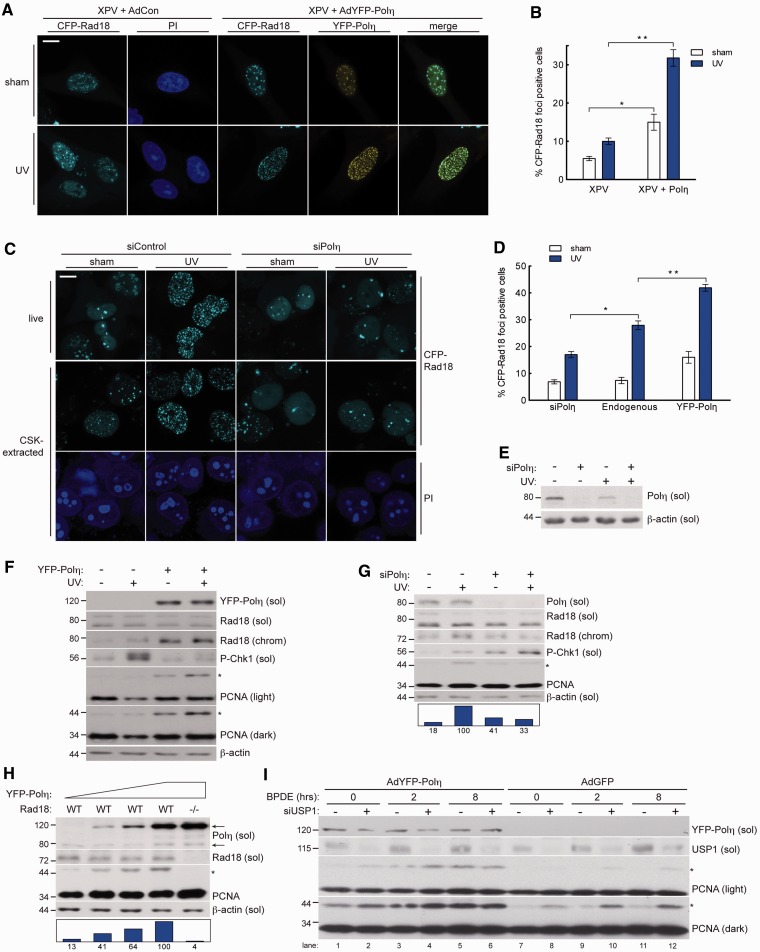
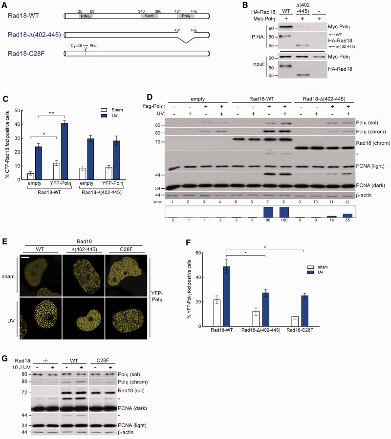
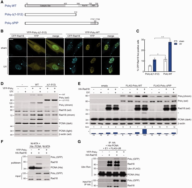
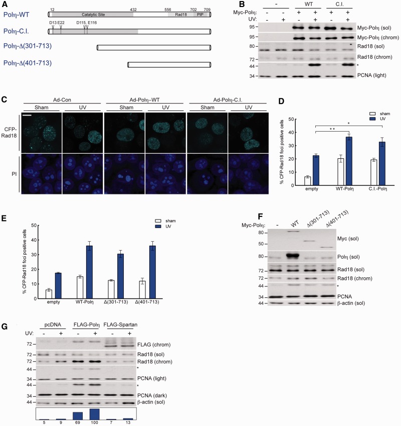
Trans-lesion DNA synthesis (TLS) is a DNA damage-tolerance mechanism that uses low-fidelity DNA polymerases to replicate damaged DNA. The inherited cancer-propensity syndrome xeroderma pigmentosum variant (XPV) results from error-prone TLS of UV-damaged DNA. TLS is initiated when the Rad6/Rad18 complex monoubiquitinates proliferating cell nuclear antigen (PCNA), but the basis for recruitment of Rad18 to PCNA is not completely understood. Here, we show that Rad18 is targeted to PCNA by DNA polymerase eta (Polη), the XPV gene product that is mutated in XPV patients. The C-terminal domain of Polη binds to both Rad18 and PCNA and promotes PCNA monoubiquitination, a function unique to Polη among Y-family TLS polymerases and dissociable from its catalytic activity. Importantly, XPV cells expressing full-length catalytically-inactive Polη exhibit increased recruitment of other error-prone TLS polymerases (Polκ and Polι) after UV irradiation. These results define a novel non-catalytic role for Polη in promoting PCNA monoubiquitination and provide a new potential mechanism for mutagenesis and genome instability in XPV individuals.


Figures

Figure 1.
Figure 1.
Polη promotes damage-induced Rad18 redistribution and PCNA monoubiquitination. (A) Representative images of CSK-extracted nuclei from CFP-Rad18-expressing XP115LO XPV cells co-infected with empty control adenovirus or adenovirus expressing YFP-Polη (at levels that restore UV tolerance—see Supplementary Figure S6) and exposed to UV (10 J/m2) or sham irradiated. Scalebar = 10 μm. (B) Quantification of CFP-Rad18 foci-positive H1299 nuclei as a percentage of CFP-Rad18-expressing cells as shown in (A); *P = 0.0001; **P = 0.001. Error bars = SEM. (C) Representative images of live (top) and CSK-extracted (bottom) nuclei from H1299 cells treated with non-targeting control siRNA (left) or siRNA targeting Polη (right) and imaged 2 h after sham or UV (10 J/m2) irradiation. Scalebar = 10 microns. (D) Quantification of CFP-Rad18 foci-positive nuclei as a percentage of CFP-Rad18-expressing cells as shown in (D). *P = 0.001; **P = 0.0003. (E) Immunoblot of fractionated lysates from H1299 cells expressing CFP-tagged Rad18 as shown in (D) and treated with non-targeting control siRNA or siRNA against Polη and then lysed 2 h after treatment with 10 J/m2 UV or sham treated. (F) Immunoblot of fractionated lysates from XP115LO XPV cells treated with empty control adenovirus or adenovirus expressing YFP-Polη at levels shown in (A) and lysed 2 h after treatment with 10 J/m2 UV. (G) Immunoblot of fractionated lysates from HDF cells treated with non-targeting control siRNA or siRNA against Polη and then lysed 2 h after treatment with 10 J/m2 UV or sham irradiation. (H) Immunoblot of fractionated lysates from HCT-116 WT cells (lanes 1–4) or RAD18−/− cells (lane 5) treated with increasing titers of YFP-Polη adenovirus and lysed 24 h post-infection. Upper and lower arrows denote YFP-tagged and endogenous Polη, respectively. (I) Immunoblot of fractionated lysates from H1299 cells expressing empty control adenovirus or adenovirus expressing YFP-Polη and treated with non-targeting control siRNA or siRNA against USP1 and then lysed at indicated times after treatment with 200 nM BPDE. On all Western blots, asterisk denotes monoubiquitinated PCNA and bar graphs represent intensity of monoubiquitinated PCNA band relative to the maximum band on each film.
Figure 2.
Figure 2.
Physical interaction between Rad18 and Polη drives efficient damage-induced PCNA ubiquitination. (A) Schematic of Rad18-WT (top); Rad18-Δ(402–445), a Polη-binding deficient mutant lacking the Polη binding domain (AA 402–445); Rad18-C28F, an E3 ligase-inactive mutant in which the RING-finger cysteine has been substituted with phenylalanine. (B) Immunoblot analysis of anti-HA immunoprecipitates from H1299 cells co-expressing HA-Rad18-WT or HA-Rad18-Δ(402–445) with Myc-Polη. (C) Quantification of CFP-Rad18 foci-positive nuclei as a percentage of H1299 cells expressing CFP-Rad18-WT or CFP-Rad18-Δ(402–445) and treated with empty adenovirus or Myc-Polη-expressing adenovirus followed by UV (10 J/m2) treatment or sham irradiation. *P = 0.095; **P = 0.0006. Error bars = SEM. (D) Immunoblots of fractionated lysates from Rad18-depleted H1299 cells that were reconstituted with siRNA-resistant Rad18-WT, Rad18-Δ(402–445), or empty vector (for control) alone or together with FLAG-Polη, followed by treatment with UV (10 J/m2) or sham-irradiation. (E) Representative images of CSK-extracted nuclei from YPF-Polη expressing H1299 cells that were depleted of endogenous Rad18 and then reconstituted with siRNA-resistant Rad18-WT, or Rad18-Δ(402–445), or Rad18-C28F and treated with UV (10 J/m2) or sham irradiated. Scalebar = 10 um. (F) Quantification of YFP-Polη foci-positive nuclei as a percentage of YFP-Polη-expressing cells in cultures complemented with Rad18-WT, Rad18-Δ(402–445), or Rad18-C28F as shown in (E). *upper P = 0.026, lower P = 0.0238. (G) Immunoblots of fractionated lysates from Rad18-depleted H1299 cells that were reconstituted with siRNA-resistant WT-Rad18, C28F-Rad18, or empty vector, and then treated with UV (10 J/m2) or sham irradiated.
Figure 3.
Figure 3.
Polη physically bridges Rad18 and PCNA to promote efficient PCNA monoubiquitination after DNA damage. (A) Schematic of Polη-WT (top); Polη-Δ(1–512), a C-terminal truncation lacking AA 513–713 (middle); and Polη-ΔPIP, full length Polη with two PIP box phenylalanines mutated to alanine (bottom). (B) Representative images of CSK-extracted nuclei from XPV cells that were co-infected with CFP-Rad18 and YFP-Polη-Δ(1–512) adenovirus (left) or YFP-Polη-WT (right) and treated with UV (10 J/m2) or sham irradiated. Scalebar = 10 um. (C) Quantification of CFP-Rad18 foci-positive nuclei as a percentage of CFP-Rad18-expressing XPV cells expressing YFP-Polη-Δ(1–512) or YFP-Polη-WT adenovirus. *upper P = 0.018; **P = 0.0001; Error bars = SEM. (D) Immunoblots of fractionated lysates from XPV cells complemented with Polη-WT or Polη-Δ(1–512) and treated with 10 J/m2 UV. (E) Immunoblots of fractionated lysates from Rad18-depleted H1299 cells that were reconstituted with siRNA-resistant Rad18-WT together with FLAG-tagged Polη-WT or Polη-ΔPIP and treated with sham or 10 J/m2 UV. (F) In vitro pulldown assay. His6-PCNA-loaded Nickel beads (or unloaded beads) were incubated with lysates from UV-irradiated H1299 cells expressing HA-Rad18 or both HA-Rad18 and YFP-Polη. (G) In vitro ubiquitination assay. HA-Rad18 complexes immunoprecipitated from UV-irradiated H1299 cells expressing HA-Rad18 alone or in combination with YFP-Polη were mixed with recombinant His6-PCNA, E1, FLAG-ubiquitin, and an ATP-regenerating system and conjugated FLAG-Ub was detected by immunoblotting with anti-FLAG antibodies.
Figure 4.
Figure 4.
Physical bridging of Rad18 and PCNA by Polη is dissociable from its DNA polymerase activity. (A) Schematic of Polη-WT (top); full-length catalytically inactive Polη, Polη-C.I, in which amino acids D13, E22, D115 and E116 are mutated to alanine; and N-terminal Polη truncation mutants, Polη-Δ(301–713) and Polη-Δ(401–713). (B) Immunoblots of fractionated lysates from Myc-Polη-WT or Myc-Polη-C.I.-expressing H1299 cells that were treated with UV (10 J/m2) or sham irradiated. (C) Representative images of CSK-extracted nuclei from H1299 cells that were co-infected with CFP-Rad18 and empty control adenovirus (left), Myc-Polη-WT (middle) or Myc-Polη-C.I. (right) and treated with UV (10 J/m2) or sham irradiated. Scalebar = 10 um. (D) Quantification of CFP-Rad18 foci-positive nuclei as a percentage of CFP-Rad18-expressing H1299 cells expressing empty control adenovirus, Myc-Polη-WT or Myc-Polη-C.I. **P = 0.0016; *P = 0.0287; Error bars = SEM. (E) Quantification of CFP-Rad18 foci-positive nuclei as a percentage of CFP-Rad18-expressing H1299 cells expressing empty control adenovirus, Myc-Polη-WT, Myc-Polη-Δ(301–713) or Myc-Polη-Δ(401–713). Error bars = SEM. (F) Immunoblot of fractionated lysates from H1299 cells expressing empty control adenovirus, Myc-Polη-WT, Myc-Polη-Δ(301–713) or Myc-Polη-Δ(401–713). (G) Immunoblot of fractionated lysates from H1299 cells expressing empty vector control, FLAG-Polη or FLAG-Spartan and lysed 2 h after treatment with 10 J/m2 UV or sham treatment.
Figure 5.
Figure 5.
High-affinity interaction with PCNA drives Polη-specific induction of PCNA monoubiquitination. (A) Immunoblot of fractionated lysates from control or Polκ-depleted H1299 cells that were lysed 2 h after treatment with UV (10 J/m2) or sham irradiation. (B) Immunoblot of fractionated lysates from H1299 cells expressing YFP-Polη or GFP-Polκ and lysed 2 h after treatment with UV (10 J/m2) or sham irradiated. (C) Sequence of the C-terminus of Polη and Polκ and the mutants used in domain-swap experiments: Polη-ΔPLTH and Polκ+PLTH. PIP-box consensus amino acids are in bold, where ψ = I/L/M; θ = Y/F. (D) Representative images of CSK-extracted nuclei from H1299 cells that were infected with GFP-Polκ-WT, GFP-Polκ+PLTH, YFP-Polη-WT or YFP-Polη-ΔPLTH and treated with 10 J/m2 UV or sham irradiated. Scalebar = 10 μm. (E) Quantification of foci-positive nuclei as a percentage of H1299 cells expressing in YFP-Polη-WT, YFP-Polη-ΔPLTH, GFP-Polκ-WT or GFP-Polκ+PLTH. *left P = 0.0001; **P = 0.0004; Error bars = SEM. (F) Immunoblot of fractionated lysates from H1299 expressing YFP-Polη-WT, YFP-Polη-ΔPLTH, GFP-Polκ-WT or GFP-Polκ+PLTH. (G) Representative images of CSK-extracted nuclei from XPV cells that were co-infected with CFP-Polι or GFP-Polκ and empty control adenovirus (left), Myc-Polη-WT (middle) or Myc-Polη-C.I. and treated with UV (10 J/m2) or sham irradiated. Scalebar = 10 µm. (H) Quantification of CFP-Polι foci-positive nuclei as a percentage of CFP-Polι-expressing XPV cells (left) and GFP-Polκ foci-positive nuclei as a percentage of GFP-Polκ-expressing XPV cells (right), after co-infection with empty control adenovirus, Myc-Polη-WT or Myc-Polη-C.I. and treatment with UV (10 J/m2) or sham irradiation. *left P = 0.0009; **P = 0.0004, *right P = 0.0022; Error bars = SEM.
Figure 6.
Figure 6.
Rad18–Polη interaction is checkpoint sensitive and p53 regulated in response to DNA damage. (A) Immunoblots of fractionated lysates from H1299 cells transfected with increasing quantities of pACCMV-Rad18-Polη fusion construct. (B) Immunoblot of fractionated lysates from HCT-116 WT or HCT-116 p53−/− cells that were UV treated (30 J/m2) and lysed at indicated times after irradiation. (C) Immunoblots of fractionated lysates of H1299 cells that were transfected with empty pcDNA as control or pcDNA-p53, followed by non-targeting control siRNA or siRNA against Polη. Cells were lysed 6 h after 10 J/m2 UV. (D) UV sensitivity of WT or p53−/− HDF incubated in 1 mM caffeine and exposed to increasing doses of UV. Cells were infected with YFP-Polη adenovirus at a dose that confers UV survival in XP115LO cells (see Supplementary Figure S6). **P = 0.0305 at 12 J/m2. **P = 0.0036 at 15 J/m2.
Figure 7.
Figure 7.
Contributions of p53 and Chk1 signalling to Polη-facilitated PCNA monoubiquitination. UV-induced p53 activity leads to transcriptional induction of Polη expression (left). RPA-coated ssDNA generated through helicase-polymerase uncoupling directly recruits Rad18 and promotes Polη-Rad18 association via Chk1 signalling (right), thereby stimulating PCNA monoubiquitination and dependent DDR pathways.

Similar articles

Cited by

References

    1. Friedberg EC. DNA damage and repair. Nature. 2003;421:436–440. - PubMed
    1. Friedberg EC, Wagner R, Radman M. Specialized DNA polymerases, cellular survival, and the genesis of mutations. Science. 2002;296:1627–1630. - PubMed
    1. Friedberg EC, Fischhaber PL, Kisker C. Error-prone DNA polymerases: novel structures and the benefits of infidelity. Cell. 2001;107:9–12. - PubMed
    1. Goodman MF. Error-prone repair DNA polymerases in prokaryotes and eukaryotes. Annu. Rev. Biochem. 2002;71:17–50. - PubMed
    1. Ohmori H, Friedberg EC, Fuchs RP, Goodman MF, Hanaoka F, Hinkle D, Kunkel TA, Lawrence CW, Livneh Z, Nohmi T, et al. The Y-family of DNA polymerases. Mol. Cell. 2001;8:7–8. - PubMed

Publication types

MeSH terms