

A case of ascending colon variceal bleeding treated with venous coil embolization
- PMID: 23345957
- PMCID: PMC3547563
- DOI: 10.3748/wjg.v19.i2.311
A case of ascending colon variceal bleeding treated with venous coil embolization
Abstract
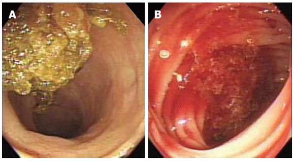
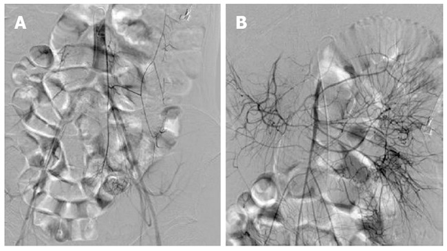
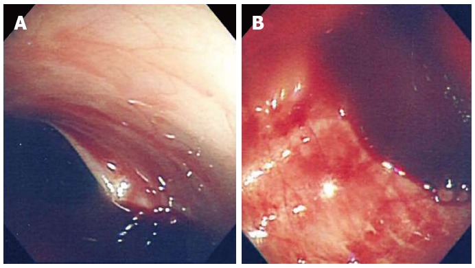
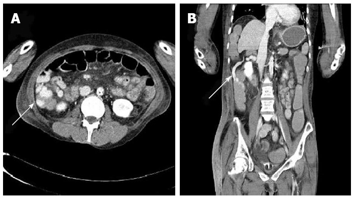
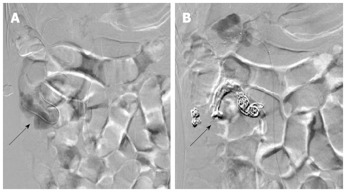
A 38-year-old female with a history of alcoholic liver cirrhosis visited our hospital with a massive hematochezia. An esophagogastroduodenoscopy did not demonstrate any bleeding source, and a colonoscopy showed a massive hemorrhage in the ascending colon but without an obvious focus. The source of the bleeding could not be found with a mesenteric artery angiography. We performed an enhanced abdominal computed tomography, which revealed a distal ascending colonic varix, and assumed that the varix was the source of the bleeding. We performed a venous coil embolization and histoacryl injection to obliterate the colon varix. The intervention appeared to be successful because the vital signs and hemoglobin laboratory data remained stable and because the hematochezia was no longer observed. We report here on a rare case of colonic variceal bleeding that was treated with venous coil embolization.
Keywords: Colon ascending; Liver cirrhosis; Therapeutic embolization; Varicose veins.
Figures
Similar articles
-
[Ascending Colon Variceal Bleeding in Cirrhotic Patient with Emergent Endoscopic Variceal Obturation with N-butyl-2-cyanoacrylate].Korean J Gastroenterol. 2018 Jul 25;72(1):37-41. doi: 10.4166/kjg.2018.72.1.37. Korean J Gastroenterol. 2018. PMID: 30049177 Korean.
-
Jejunal variceal bleeding successfully treated with percutaneous coil embolization.J Korean Med Sci. 2012 Mar;27(3):321-4. doi: 10.3346/jkms.2012.27.3.321. Epub 2012 Feb 23. J Korean Med Sci. 2012. PMID: 22379346 Free PMC article.
-
Histoacryl injection for treatment of varices in the ascending colon.Endoscopy. 2016;48 Suppl 1:E285. doi: 10.1055/s-0042-114427. Epub 2016 Sep 14. Endoscopy. 2016. PMID: 27626210 No abstract available.
-
Endoscopic band ligation for transverse colonic variceal bleeding: case report and review of the literature.Ann Saudi Med. 2020 May-Jun;40(3):255-258. doi: 10.5144/0256-4947.2020.255. Epub 2020 Jun 4. Ann Saudi Med. 2020. PMID: 32493047 Free PMC article. Review.
-
Bleeding from a duodenal varix: a unique case of variceal hemostasis achieved using EUS-guided placement of an embolization coil and cyanoacrylate.J Clin Gastroenterol. 2014 Apr;48(4):362-4. doi: 10.1097/MCG.0000000000000004. J Clin Gastroenterol. 2014. PMID: 24518801 Review.
Cited by
-
Lower GI Bleeding: An Update on Incidences and Causes.Clin Colon Rectal Surg. 2020 Jan;33(1):28-34. doi: 10.1055/s-0039-1695035. Epub 2019 Nov 11. Clin Colon Rectal Surg. 2020. PMID: 31915423 Free PMC article. Review.
-
A Unique Presentation of Familial Idiopathic Colonic Varices.ACG Case Rep J. 2023 Nov 2;10(11):e01185. doi: 10.14309/crj.0000000000001185. eCollection 2023 Nov. ACG Case Rep J. 2023. PMID: 37928226 Free PMC article.
-
Rupture of ectopic varices of the ascending colon occurring after pancreatic cancer surgery: A case report and literature review.DEN Open. 2023 Jul 10;4(1):e255. doi: 10.1002/deo2.255. eCollection 2024 Apr. DEN Open. 2023. PMID: 37441155 Free PMC article.
-
Percutaneous Transhepatic Embolization of a Bleeding Colic Vein in a Cirrhotic Patient With Massive Hematochezia: A Case Report and Literature Review.Cureus. 2022 Jun 7;14(6):e25736. doi: 10.7759/cureus.25736. eCollection 2022 Jun. Cureus. 2022. PMID: 35812565 Free PMC article.
-
Massive Cecal Variceal Hemorrhage Treated with Transjugular Intrahepatic Portosystemic Shunt with Right Colic Vein and Ileocolic Vein Embolization.Cureus. 2019 Apr 5;11(4):e4392. doi: 10.7759/cureus.4392. Cureus. 2019. PMID: 31223550 Free PMC article.
References
-
- Kim HU, Her KH, Kim SH, Kim BS, Kang YJ, Lee J, Kim KS. A case of variceal bleeding of the ascending colon associated with alcoholic liver cirrhosis. Korean J Med. 2008;75:215–220.
-
- Edwards EA. Functional anatomy of the porta-systemic communications. AMA Arch Intern Med. 1951;88:137–154. - PubMed
-
- Francois F, Tadros C, Diehl D. Pan-colonic varices and idiopathic portal hypertension. J Gastrointestin Liver Dis. 2007;16:325–328. - PubMed
Publication types
MeSH terms
Substances
LinkOut - more resources
Full Text Sources
Other Literature Sources
Medical
