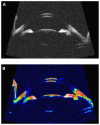
Anterior segment biometry using ultrasound biomicroscopy and the Artemis-2 very high frequency ultrasound scanner
- PMID: 23345968
- PMCID: PMC3551458
- DOI: 10.2147/OPTH.S39463
Anterior segment biometry using ultrasound biomicroscopy and the Artemis-2 very high frequency ultrasound scanner
Abstract
Purpose: To compare the precision of anterior chamber angle (ACA) and anterior chamber depth (ACD) measurements taken with ultrasound biomicroscopy (UBM) and the Artemis-2 Very High Frequency Ultrasound Scanner (VHFUS) in normal subjects.
Design: Prospective study.
Methods: We randomly selected one eye from each of 59 normal subjects in this study. Two subjects dropped out of the study; the associated data were excluded from analysis. ACA and ACD measurements were obtained using the VHFUS and the UBM. The results were compared statistically using repeated-measures analysis of variance for the intraobserver repeatability, unpaired t-test, and limits of agreement.
Results: The average ACA values for the UBM and the VHFUS (±standard deviation) were 41.83° ± 5.03° and 33.36° ± 6.03°, respectively. The average ACD values were 2.96 ± 0.34 mm and 2.87 ± 0.31 mm. The intraobserver repeatability analysis of variance P-values for ACA and ACD measurements using UBM were 0.10 and 0.68, respectively; for the Artemis-2 VHFUS, the respective values were 0.68 and 0.09. The difference in ACA measurements was statistically significant (t = 8.41; P < 0.0001), while the difference in ACD values was not (t = 1.51; P < 0.13). The mean ACA difference was 8.50° ± 2.50°, and the limits of agreement were +13.30° to -3.60°. The mean ACD difference was 0.09 ± 0.27 mm, and the limits of agreement ranged from 0.61 mm to -0.43 mm. The mean difference percentage of ACD was 3.1% for both instruments.
Conclusion: In case of the ACD, both instruments can be used interchangeably; however, with the ACA instruments, they cannot be used interchangeably.
Keywords: Artemis-2 VHF scanner; anterior chamber angle; anterior chamber depth; normal eyes; ultrasound biomicroscope.
Figures


References
-
- Sihota R, Lakshimaiah NC, Agrawal HC, Pandey RM, Titiyal JS. Ocular parameters in the subgroups of angle closure glaucoma. Clin Experiment Ophthalmol. 2000;28(4):253–258. - PubMed
-
- Baikoff G, Lutun E, Ferraz C, Wei J. Static and dynamic analysis of the anterior segment with optical coherence tomography. J Cataract Refract Surg. 2004;30(9):1843–1850. - PubMed
-
- Radhakrishnan S, Goldsmith J, Huang D, et al. Comparison of optical coherence tomography and ultrasound biomicroscopy for detection of narrow anterior chamber angles. Arch Ophthalmol. 2005;123(8):1053–1059. - PubMed
LinkOut - more resources
Full Text Sources
Other Literature Sources

