. 2013:8:325-36.
doi: 10.2147/IJN.S36711. Epub 2013 Jan 18.

Evaluation of gold nanotracers to track adipose-derived stem cells in a PEGylated fibrin gel for dermal tissue engineering applications

Affiliations

Evaluation of gold nanotracers to track adipose-derived stem cells in a PEGylated fibrin gel for dermal tissue engineering applications

Eunna Chung et al. Int J Nanomedicine. 2013.

Abstract

Evaluating the regenerative capacity of a tissue-engineered device in a noninvasive and synchronous manner is critical to determining the mechanisms for success in clinical applications. In particular, directly tracking implanted cells in a three-dimensional (3D) scaffold is desirable in that it enables the monitoring of cellular activity in a specific and localized manner. The authors' group has previously demonstrated that the PEGylation of fibrin results in a 3D scaffold that supports morphologic and phenotypic changes in mesenchymal stem cells that may be advantageous in wound healing applications. Recently, the authors have evaluated adipose-derived stem cells (ASCs) as a mesenchymal cell source to regenerate skin and blood vessels due to their potential for proliferation, differentiation, and production of growth factors. However, tracking and monitoring ASCs in a 3D scaffold, such as a PEGylated fibrin gel, have not yet been fully investigated. In the current paper, nanoscale gold spheres (20 nm) as cell tracers for ASCs cultured in a PEGylated fibrin gel were evaluated. An advanced dual-imaging modality combining ultrasound and photoacoustic imaging was utilized to monitor rat ASCs over time. The ASCs took up gold nanotracers and could be detected up to day 16 with high sensitivity using photoacoustic imaging. There were no detrimental effects on ASC morphology, network formation, proliferation, and protein expression/secretion (ie, smooth muscle α-actin, vascular endothelial growth factor, matrix metalloproteinase-2, and matrix metalloproteinase-9) associated with gold nanotracers. Therefore, utilization of gold nanotracers can be an effective strategy to monitor the regenerative process of a stem cell source in a 3D gel for vascular and dermal tissue engineering applications.

Keywords: adipose-derived stem cells; angiogenesis; fibrin; gold nanoparticles; tissue engineering; ultrasound and photoacoustic imaging.


Figures
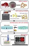
Figure 1
Figure 1
Schematic workflow for gold nanotracer-mediated tracking of adipose-derived stem cells in a three-dimensional PEGylated fibrin gel. Abbreviations: 3D, three-dimensional; ADSC, adipose-derived stem cell; GNT, gold nanotracer; SC-PEG-SC, succinimidyl glutarate polyethylene glycol.
Figure 2
Figure 2
(A and B) Transmission electron microscope images of gold nanotracer-loaded adipose-derived stem cells. Red arrows indicate aggregated 20 nm gold nanotracers in the cytoplasm. (C) Ultraviolet-visible spectrum of gold nanotracers. (D) Ultraviolet-visible spectrum of cells and gold nanotracer-loaded cells. Abbreviations: GNT, gold nanotracer; a.u., arbitrary unit.
Figure 3
Figure 3
Bright field, phase contrast, and dark field optical images of monolayer adipose-derived stem cells with and without gold nanotracers on day 1 and day 5. Abbreviations: DAPI, 4′,6-diamidino-2-phenylindole; GNT, gold nanotracer.
Figure 4
Figure 4
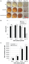
Gel degradation of a PEGylated fibrin gel over time. (A) Digital camera photos of gels on days one, four, eight, and 16. (B) Quantification of the surface area of the gels from the front view. (C) Proliferation of gold nanotracer-loaded adipose-derived stem cells in the PEGylated fibrin gel. Notes: A methyl tetrazolium salt assay was used to derive cell numbers in adipose-derived stem cells with and without gold nanotracers in the gel over time. The data levels that do not share the same letters are significantly different (P < 0.05). Abbreviations: ADSC, adipose-derived stem cell; GNT, gold nanotracer.
Figure 5
Figure 5
Dual ultrasound and photoacoustic imaging of adipose-derived stem cell-cultured PEGylated fibrin gels. (A) Reconstructed morphology of the fibrin gels (top two) and ultrasound/photoacoustic images at the center cross-section of the gels (bottom two). (B) Photoacoustic signal intensity per volume corresponding to the photoacoustic images in Figure 5A. (C) Fibrin gel volume. (D) Total photoacoustic signal intensity. Note: The data levels that do not share the same letters are significantly different (P < 0.05). Abbreviations: 3D, three-dimensional; PA, photoacoustic; US, ultrasound; a.u., arbitrary unit.
Figure 6
Figure 6
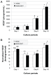
Vascular endothelial growth factor secretion from adipose-derived stem cells in a PEGylated gel. (A) Total amount of vascular endothelial growth factor secretion in an individual culture system. (B) Normalized amount of vascular endothelial growth factor secretion per cell. Note: The data levels that do not share the same letters are significantly different (P < 0.05). Abbreviations: ADSC, adipose-derived stem cell; GNT, gold nanotracer; VEGF, vascular endothelial growth factor.
Figure 7
Figure 7
Matrix metalloproteinase-2 secretion from adipose-derived stem cells in a PEGylated gel. (A) Total amount of matrix metalloproteinase-2 secretion in a individual culture system. (B) Normalized amount of matrix metalloproteinase-2 secretion per cell. Note: The data levels that do not share the same letters are significantly different (P < 0.05). Abbreviations: ADSC, adipose-derived stem cell; GNT, gold nanotracer; MMP-2, matrix metalloproteinase-2.
Figure 8
Figure 8
Matrix metalloproteinase-9 secretion from adipose-derived stem cells in a PEGylated gel. (A) Total amount of matrix metalloproteinase-9 secretion in an individual culture system. (B) Normalized amount of matrix metalloproteinase-9 secretion per cell. Note: The data levels that do not share the same letters are significantly different (P < 0.05). Abbreviations: ADSC, adipose-derived stem cell; GNT, gold nanotracer; MMP-9, matrix metalloproteinase-9.
Figure 9
Figure 9
Immunofluorescence staining of smooth muscle α-actin over time (days one, four, eight, and 16). Abbreviations: ADSC, adipose-derived stem cell; DAPI, 4′,6-diamidino-2-phenylindole; GNT, gold nanotracer; SMA, smooth muscle α-actin.

Similar articles

Cited by

References

    1. Espandar L, Bunnell B, Wang GY, Gregory P, McBride C, Moshirfar M. Adipose-derived stem cells on hyaluronic acid-derived scaffold: a new horizon in bioengineered cornea. Arch Ophthalmol. 2012;130(2):202–208. - PubMed
    1. Nam SY, Ricles LM, Suggs LJ, Emelianov SY. In vivo ultrasound and photoacoustic monitoring of mesenchymal stem cells labeled with gold nanotracers. PLoS One. 2012;7(5):e37267. - PMC - PubMed
    1. Lalande C, Miraux S, Derkaoui SM, et al. Magnetic resonance imaging tracking of human adipose derived stromal cells within three-dimensional scaffolds for bone tissue engineering. Eur Cell Mater. 2011;21:341–354. - PubMed
    1. Kim K, Jeong CG, Hollister SJ. Non-invasive monitoring of tissue scaffold degradation using ultrasound elasticity imaging. Acta Biomater. 2008;4(4):783–790. - PMC - PubMed
    1. Kreitz S, Dohmen G, Hasken S, Schmitz-Rode T, Mela P, Jockenhoevel S. Nondestructive method to evaluate the collagen content of fibrin-based tissue engineered structures via ultrasound. Tissue Eng Part C Methods. 2011;17(10):1021–1026. - PubMed

Publication types

MeSH terms