

Evaluation of gold nanotracers to track adipose-derived stem cells in a PEGylated fibrin gel for dermal tissue engineering applications
- PMID: 23345978
- PMCID: PMC3551459
- DOI: 10.2147/IJN.S36711
Evaluation of gold nanotracers to track adipose-derived stem cells in a PEGylated fibrin gel for dermal tissue engineering applications
Abstract
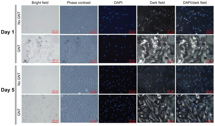
Evaluating the regenerative capacity of a tissue-engineered device in a noninvasive and synchronous manner is critical to determining the mechanisms for success in clinical applications. In particular, directly tracking implanted cells in a three-dimensional (3D) scaffold is desirable in that it enables the monitoring of cellular activity in a specific and localized manner. The authors' group has previously demonstrated that the PEGylation of fibrin results in a 3D scaffold that supports morphologic and phenotypic changes in mesenchymal stem cells that may be advantageous in wound healing applications. Recently, the authors have evaluated adipose-derived stem cells (ASCs) as a mesenchymal cell source to regenerate skin and blood vessels due to their potential for proliferation, differentiation, and production of growth factors. However, tracking and monitoring ASCs in a 3D scaffold, such as a PEGylated fibrin gel, have not yet been fully investigated. In the current paper, nanoscale gold spheres (20 nm) as cell tracers for ASCs cultured in a PEGylated fibrin gel were evaluated. An advanced dual-imaging modality combining ultrasound and photoacoustic imaging was utilized to monitor rat ASCs over time. The ASCs took up gold nanotracers and could be detected up to day 16 with high sensitivity using photoacoustic imaging. There were no detrimental effects on ASC morphology, network formation, proliferation, and protein expression/secretion (ie, smooth muscle α-actin, vascular endothelial growth factor, matrix metalloproteinase-2, and matrix metalloproteinase-9) associated with gold nanotracers. Therefore, utilization of gold nanotracers can be an effective strategy to monitor the regenerative process of a stem cell source in a 3D gel for vascular and dermal tissue engineering applications.
Keywords: adipose-derived stem cells; angiogenesis; fibrin; gold nanoparticles; tissue engineering; ultrasound and photoacoustic imaging.
Figures


References
-
- Espandar L, Bunnell B, Wang GY, Gregory P, McBride C, Moshirfar M. Adipose-derived stem cells on hyaluronic acid-derived scaffold: a new horizon in bioengineered cornea. Arch Ophthalmol. 2012;130(2):202–208. - PubMed
-
- Lalande C, Miraux S, Derkaoui SM, et al. Magnetic resonance imaging tracking of human adipose derived stromal cells within three-dimensional scaffolds for bone tissue engineering. Eur Cell Mater. 2011;21:341–354. - PubMed
-
- Kreitz S, Dohmen G, Hasken S, Schmitz-Rode T, Mela P, Jockenhoevel S. Nondestructive method to evaluate the collagen content of fibrin-based tissue engineered structures via ultrasound. Tissue Eng Part C Methods. 2011;17(10):1021–1026. - PubMed
Publication types
MeSH terms
Substances
Grants and funding
LinkOut - more resources
Full Text Sources
Other Literature Sources
Medical
Miscellaneous
