Review
. 2013:8:337-50.
doi: 10.2147/IJN.S38635. Epub 2013 Jan 18.

Techniques for fabrication and construction of three-dimensional scaffolds for tissue engineering

Affiliations
Review

Techniques for fabrication and construction of three-dimensional scaffolds for tissue engineering

Tingli Lu et al. Int J Nanomedicine. 2013.

Abstract

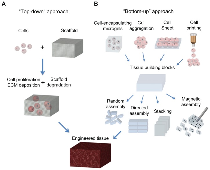
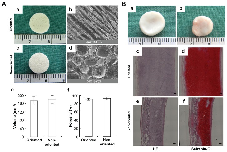
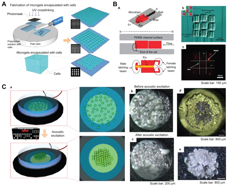
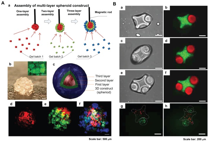
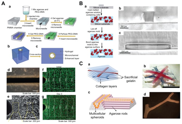
Three-dimensional biomimetic scaffolds have widespread applications in biomedical tissue engineering because of their nanoscaled architecture, eg, nanofibers and nanopores, similar to the native extracellular matrix. In the conventional "top-down" approach, cells are seeded onto a biocompatible and biodegradable scaffold, in which cells are expected to populate in the scaffold and create their own extracellular matrix. The top-down approach based on these scaffolds has successfully engineered thin tissues, including skin, bladder, and cartilage in vitro. However, it is still a challenge to fabricate complex and functional tissues (eg, liver and kidney) due to the lack of vascularization systems and limited diffusion properties of these large biomimetic scaffolds. The emerging "bottom-up" method may hold great potential to address these challenges, and focuses on fabricating microscale tissue building blocks with a specific microarchitecture and assembling these units to engineer larger tissue constructs from the bottom up. In this review, state-of-the-art methods for fabrication of three-dimensional biomimetic scaffolds are presented, and their advantages and drawbacks are discussed. The bottom-up methods used to assemble microscale building blocks (eg, microscale hydrogels) for tissue engineering are also reviewed. Finally, perspectives on future development of the bottom-up approach for tissue engineering are addressed.

Keywords: bottom-up; extracellular matrix scaffolds; three-dimensional; tissue engineering.


Figures

Figure 1
Figure 1
Schematic of “top-down” and “bottom-up” approaches for tissue engineering. (A) In the top-down approach, cells are seeded on a biocompatible and biodegradable scaffold and are expected to populate in the scaffold and create their own extracellular matrix. (B) In the bottom-up approach, various methods are utilized for generating tissue building blocks and these units can be engineered into large tissue constructs via multiple assembling methods. Abbreviation: ECM, extracellular matrix.
Figure 2
Figure 2
Electrospinning technique for three-dimensional biomimetic scaffold fabrication. (A) Schematic of electrospinning technique and (B) scanning electron microscopic images of collagen nanofibers fabricated by electrospinning.
Figure 3
Figure 3
Scanning electron micrographs of nanostructured scaffolds fabricated by phase-separation and freeze-drying. (A and B) Images of chitosan nanofibers prepared by phase-separation at 0.05% (w/v) chitosan, 0.025% (w/v), and liquid nitrogen (A, scale bar 10 μm; B, scale bar 2 μm). Images of porous scaffolds prepared by freeze-drying under 6.5 mbar (C) and 0.1 mbar (D) freeze-drying pressure (scale bar, 50 μm).
Figure 4
Figure 4
Self-assembly method for fabrication of three-dimensional scaffolds. (A) Schematic representation of filamentous nanostructures surrounding cells for signaling events. (B) (a) Chemical structure and (b) molecular model of peptide amphiphiles. Color scheme: C, black; H, white; O, red; N, blue; P, cyan; S, yellow. (c) Schematic for the self-assembly of peptide amphiphiles into a cylindrical micelle. (C) (a and b) Transmission electron microscopic images of self-assembled nanofibers before covalent capture. (a) Scale bar 50 nm and (b) scale bar 200 nm. (c and d) Transmission electron microscopic images of self-assembled nanofibers after oxidative crosslinking (c, scale bar 100 nm; d, scale bar 25 nm).
Figure 5
Figure 5
Three-dimensional biomimetic scaffolds for cartilage tissue engineering. (A) Actual and scanning electron micrographs of oriented (a and b) and nonoriented scaffolds (c and d) showing the volume and porosity of each scaffold (e and f). (B) In vivo fate of in vitro fabricated cartilage: grow view (a and b), histology and immunohistochemistry; the specimen in the oriented group shows typical and homogeneous cartilage structure with abundant cartilage-specific extracellular matrix and a large size in thickness (c and d); the specimen in the nonoriented group shows relatively heterogeneous cartilage structure with a smaller thickness (e and f). Scale bar 100 μm.
Figure 6
Figure 6
Fabrication and assembly process for microgels via railed microfluidic and acoustic field. (A) Schematic of photolithography methods used to fabricate microgels. (B) Railed microfluidic assembly of microgels, (a) schematic of a microtrain, (b) assembled 3 × 3 matrix with two different living cells using cell-encapsulating thylene glycol diacrylate solution, and (c) fluorescent images of cell-encapsulating microgels. Scale bar 150 μm. (C) (a) Schematic of acoustic assembly of microgels; (b and c) before and after acoustic excitation, single-layer formation (200 μm × 200 μm microgels, scale bar 200 μm); (d) new microgels were introduced onto a single layer to generate a double-layered structure (scale bar 200 μm); (e) multilayered structure using layer-to-layer approach (scale bar 200 μm).
Figure 7
Figure 7
Magnetic and surface tension assembly of microgels. (A) (a) Schematic of magnetic assembly of microgels; (b) magnified image of the assembled single-layer construct (scale bar 500 μm); (c and f) merged fluorescent images of three-layer spheroids; first layer gels are stained with rhodamine B (d); second layer gels are stained with FITC-dextran (e); and third layer gels are stained with 1, 1, 4, 4-tetraphenyl-1,3-butadiene (f). Scale bar 500 μm. (B) Fluorescence images of lock-and-key assemblies with one, two, and three rods per cross (a to f); fluorescence images of assembled microgels containing green-stained and red-stained cells (g and h). Scale bar 200 μm.
Figure 8
Figure 8
Microfluidic hydrogels for vascularization. (A) (a) Schematic representation of mechanically enhanced microfluidic hydrogel fabrication; (b) three-dimensional structure and (c) cross-sectional view; (d and e) hydrogels with enhanced microchannel wall. (d) Scale bar 500 μm; (e) scale bar 200 μm. (f and g) Fluorescent images of live-dead cells following the fabrication process (f) and after 3 days of perfusion culture (g). Scale bar 500 μm. (B) (a) Schematic of fabrication process for microfluidic cell-encapsulating hydrogels (b and c) cross-sectional images of agarose constructs with channel size (b) 50 μm × 70 μm and (c) 1 mm × 150 μm. (a) Scale bar 50 μm and (b) scale bar 250 μm. (C) (a) Multilayered collagen scaffold with microfluidic microchannels; (b) utilizing printed gelatin networks as a sacrificial element. Scale bar 5 mm. (c and d) Designed post fusion pattern of branched constructs using agarose rods as filling material.

Similar articles

Cited by

References

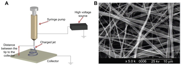
    1. Chung SW, Ingle NP, Montero GA, Kim SH, King MW. Bioresorbable elastomeric vascular tissue engineering scaffolds via melt spinning and electrospinning. Acta Biomater. 2010;6:1958–1967. - PubMed
    1. Lao LH, Wang YJ, Zhu Y, Zhang YY, Gao CY. Poly(lactide-co-glycolide)/hydroxyapatite nanofibrous scaffolds fabricated by electrospinning for bone tissue engineering. J Mater Sci Mater Med. 2011;22:1873–1884. - PubMed
    1. Soliman S, Pagliari S, Rinaldi A, et al. Multiscale three-dimensional scaffolds for soft tissue engineering via multimodal electrospinning. Acta Biomater. 2010;6:1227–1237. - PubMed
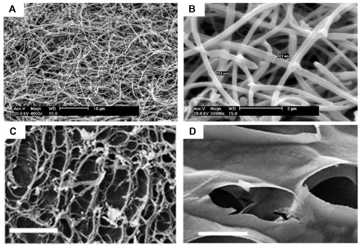
    1. Blaker JJ, Knowles JC, Day RM. Novel fabrication techniques to produce microspheres by thermally induced phase separation for tissue engineering and drug delivery. Acta Biomater. 2008;4:264–272. - PubMed
    1. Budyanto L, Goh YQ, Ooi CP. Fabrication of porous poly(L-lactide) (PLLA) scaffolds for tissue engineering using liquid-liquid phase separation and freeze extraction. J Mater Sci Mater Med. 2009;20:105–111. - PubMed

Publication types