. 2013 Jan 18:6:70.
doi: 10.3389/fncel.2012.00070. eCollection 2012.

Regulation of endogenous neural stem/progenitor cells for neural repair-factors that promote neurogenesis and gliogenesis in the normal and damaged brain

Affiliations

Regulation of endogenous neural stem/progenitor cells for neural repair-factors that promote neurogenesis and gliogenesis in the normal and damaged brain

Kimberly J Christie et al. Front Cell Neurosci. .

Abstract

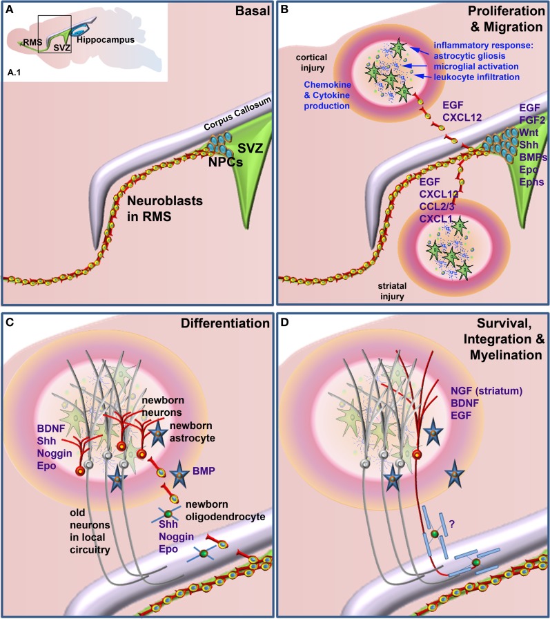
Neural stem/precursor cells in the adult brain reside in the subventricular zone (SVZ) of the lateral ventricles and the subgranular zone (SGZ) of the dentate gyrus in the hippocampus. These cells primarily generate neuroblasts that normally migrate to the olfactory bulb (OB) and the dentate granule cell layer respectively. Following brain damage, such as traumatic brain injury, ischemic stroke or in degenerative disease models, neural precursor cells from the SVZ in particular, can migrate from their normal route along the rostral migratory stream (RMS) to the site of neural damage. This neural precursor cell response to neural damage is mediated by release of endogenous factors, including cytokines and chemokines produced by the inflammatory response at the injury site, and by the production of growth and neurotrophic factors. Endogenous hippocampal neurogenesis is frequently also directly or indirectly affected by neural damage. Administration of a variety of factors that regulate different aspects of neural stem/precursor biology often leads to improved functional motor and/or behavioral outcomes. Such factors can target neural stem/precursor proliferation, survival, migration and differentiation into appropriate neuronal or glial lineages. Newborn cells also need to subsequently survive and functionally integrate into extant neural circuitry, which may be the major bottleneck to the current therapeutic potential of neural stem/precursor cells. This review will cover the effects of a range of intrinsic and extrinsic factors that regulate neural stem/precursor cell functions. In particular it focuses on factors that may be harnessed to enhance the endogenous neural stem/precursor cell response to neural damage, highlighting those that have already shown evidence of preclinical effectiveness and discussing others that warrant further preclinical investigation.

Keywords: gliogenesis; ischemic stroke; neural precursor cell (NPC); neural stem cell (NSC); neural stem/precursor cell (NSPC); neurogenesis; subventricular zone (SVZ); traumatic brain injury.


Figures

Figure 1
Figure 1
Factors that when administered to the brain following neural damage regulate neural precursor cell responses. (A) Basal, non-damage conditions. Inset (A.1) depicts the two main neurogenic regions of the adult rodent brain, the subventricular zone (SVZ) and the hippocampus. The enlarged area bounded by the box in (A.1) depicts the general structure of the SVZ, which contains proliferative neural precursor cells (NPCs) that differentiate into neuroblasts that then migrate along the rostral migratory stream (RMS) to the olfactory bulb. Following neural injury (B) the inflammatory response induces astrocytic gliosis, microglial activation, and leukocyte infiltration from the periphery. Together, these produce a variety of chemokines, cytokines and other factors that can lead to the proliferation of the NPCs in the SVZ and redirect their migration to sites of damage. Once at or near the site of damage (C) these cells can differentiate into astrocytes, oligodendrocytes and astrocytes, depending upon which factors are administered. Unlike the normal predominance of neuronal differentiation under basal conditions, following injury SVZ NSPCs can generate neurons, astrocytes and/or oligodendrocytes. (D) Following differentiation cells need to survive and appropriately integrate into existing circuitry. This is a large bottleneck in harnessing the therapeutic potential of NSPCs, as the majority of newborn neurons die within a few weeks of their generation. For correct function, many new neurons also need to be appropriately myelinated although at present, few if any factors are known that enhance the final myelination stage of oligodendrocyte maturation following neural damage. Factors that have been shown to regulate the various stages of the NPC response when administered following neural injury are listed in blue text.

Similar articles

Cited by

References

    1. Aberg M. A., Aberg N. D., Hedbacker H., Oscarsson J., Eriksson P. S. (2000). Peripheral infusion of IGF-I selectively induces neurogenesis in the adult rat hippocampus. J. Neurosci. 20, 2896–2903 - PMC - PubMed
    1. Aberg N. D., Johansson I., Aberg M. A., Lind J., Johansson U. E., Cooper-Kuhn C. M., et al. (2009). Peripheral administration of GH induces cell proliferation in the brain of adult hypophysectomized rats. J. Endocrinol. 201, 141–150 10.1677/JOE-08-0495 - DOI - PubMed
    1. Adachi K., Mirzadeh Z., Sakaguchi M., Yamashita T., Nikolcheva T., Gotoh Y., et al. (2007). Beta-catenin signaling promotes proliferation of progenitor cells in the adult mouse subventricular zone. Stem Cells 25, 2827–2836 10.1634/stemcells.2007-0177 - DOI - PubMed
    1. Agasse F., Bernardino L., Kristiansen H., Christiansen S. H., Ferreira R., Silva B., et al. (2008). Neuropeptide Y promotes neurogenesis in murine subventricular zone. Stem Cells 26, 1636–1645 10.1634/stemcells.2008-0056 - DOI - PubMed
    1. Agis-Balboa R. C., Arcos-Diaz D., Wittnam J., Govindarajan N., Blom K., Burkhardt S., et al. (2011). A hippocampal insulin-growth factor 2 pathway regulates the extinction of fear memories. EMBO J. 30, 4071–4083 10.1038/emboj.2011.293 - DOI - PMC - PubMed

LinkOut - more resources