

Familial Transthyretin Amyloidosis with Variant Asp38Ala Presenting with Orthostatic Hypotension and Chronic Diarrhea
- PMID: 23346293
- PMCID: PMC3542517
- DOI: 10.4250/jcu.2012.20.4.209
Familial Transthyretin Amyloidosis with Variant Asp38Ala Presenting with Orthostatic Hypotension and Chronic Diarrhea
Abstract
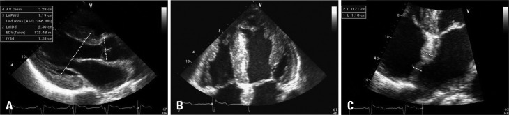
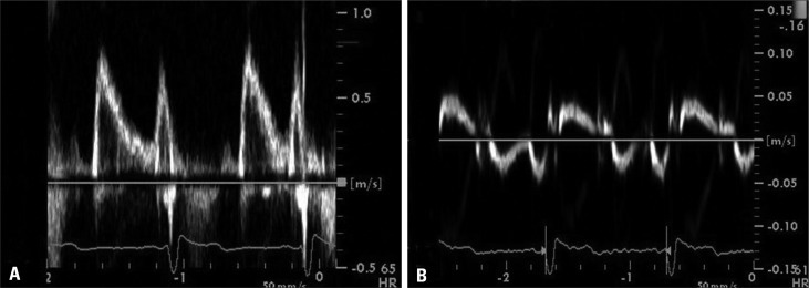
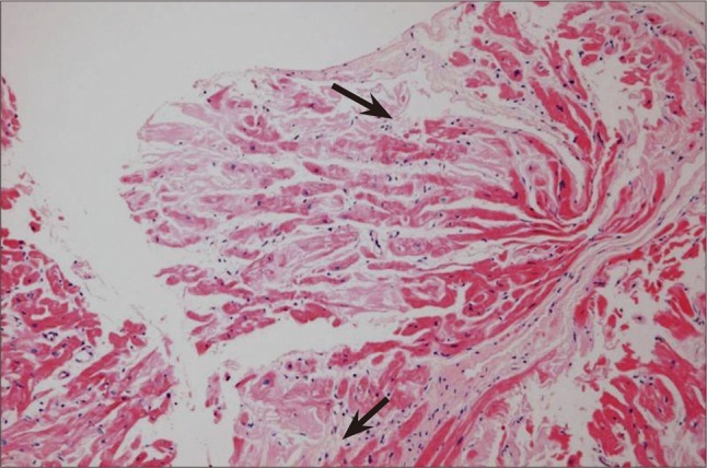
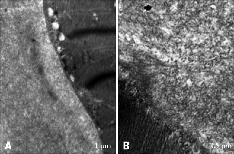
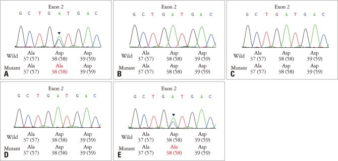
A 53-year-old man complained of orthostatic, non-rotating dizziness, and chronic watery diarrhea of several years duration. His nerve-conduction velocity test revealed peripheral sensory-motor polyneuropathy and he showed an autonomic function abnormality. Echocardiographic examination showed ventricular and atrial wall thickening with a granular "sparkling" appearance. Left ventricular systolic function was preserved but pseudonormal diastolic dysfunction was present. Coronary angiography showed normal coronary arteries and an endomyocardial biopsy revealed lesions consistent with cardiac amyloidosis. Colonoscopic biopsy also revealed the deposition of amyloid fibrils. Gene analysis found the transthyretin variant Asp38Ala. His son had same mutation, but three daughters did not. In conclusion, we report a case of familial transthyretin amyloidosis with Asp38Ala.
Keywords: Amyloidosis; Asp38Ala; Orthostatic hypotension; Polyneuropathy; Transthyretin.
Figures
References
-
- Falk RH, Comenzo RL, Skinner M. The systemic amyloidoses. N Engl J Med. 1997;337:898–909. - PubMed
-
- Westermark P, Benson MD, Buxbaum JN, Cohen AS, Frangione B, Ikeda S, Masters CL, Merlini G, Saraiva MJ, Sipe JD. Amyloid fibril protein nomenclature -- 2002. Amyloid. 2002;9:197–200. - PubMed
-
- Westermark P, Benson MD, Buxbaum JN, Cohen AS, Frangione B, Ikeda S, Masters CL, Merlini G, Saraiva MJ, Sipe JD. A primer of amyloid nomenclature. Amyloid. 2007;14:179–183. - PubMed
-
- Ando Y, Nakamura M, Araki S. Transthyretin-related familial amyloidotic polyneuropathy. Arch Neurol. 2005;62:1057–1062. - PubMed
-
- Falk RH, Dubrey SW. Amyloid heart disease. Prog Cardiovasc Dis. 2010;52:347–361. - PubMed
Publication types
LinkOut - more resources
Full Text Sources
Research Materials
