

Endoscope-Assisted Trans-Sphenoidal Approach for Treatment of Sternberg's Canal
- PMID: 23346329
- PMCID: PMC3550425
- DOI: 10.3340/jkns.2012.52.6.555
Endoscope-Assisted Trans-Sphenoidal Approach for Treatment of Sternberg's Canal
Abstract
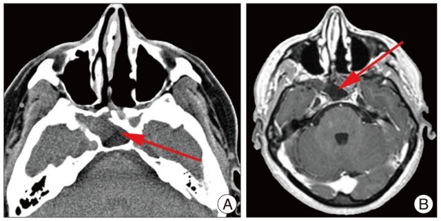
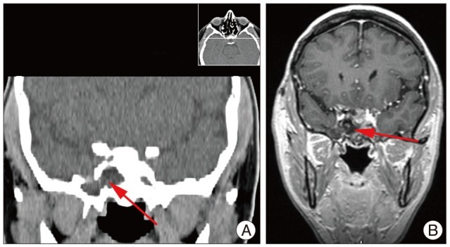
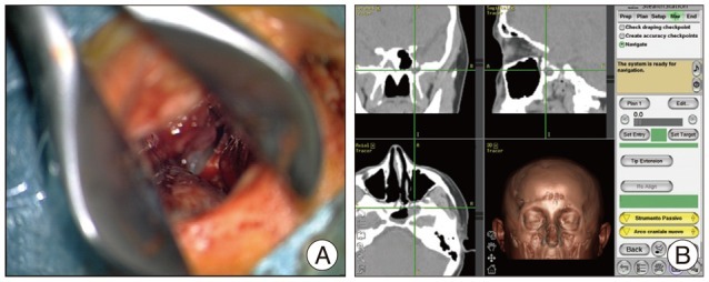
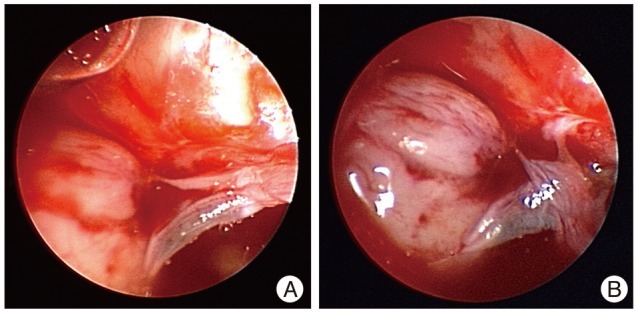
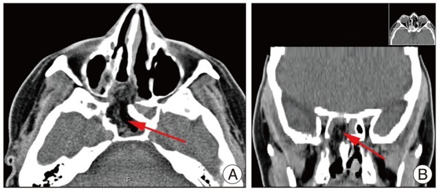
We report an uncommon case of a 45-year-old woman who presented with spontaneous rhinorrhea. A computed tomography (CT) scan of the head revealed an abnormally large sphenoid sinus associated with a parasellar bony defect (Sternberg's canal) through which magnetic resonance imaging could detect an encephalocele of the right temporal lobe. An endoscope-assisted trans-sphenoidal approach was performed and, with the aid of image guided surgery, reduction of the encephalocele was obtained and followed by surgical repair of the dural and bony defects. The postoperative course was uneventful and the cerebrospinal fluid fistula was closed as confirmed by the postoperative CT scan and by the absence of rhinorrhea. After three years of monitoring the patient remained asymptomatic.
Keywords: Cerebrospinal fluid; Endoscope-assisted procedures; Sternberg's canal.
Figures
Similar articles
-
Spontaneous sphenoid sinus cerebrospinal fluid leak and meningoencephalocele - are they due to patent Sternberg's canal?Wideochir Inne Tech Maloinwazyjne. 2015 Jul;10(2):347-58. doi: 10.5114/wiitm.2014.47097. Epub 2014 Dec 3. Wideochir Inne Tech Maloinwazyjne. 2015. PMID: 26240642 Free PMC article.
-
Sternberg's canal as a cause of encephalocele within the lateral recess of the sphenoid sinus: A report of two cases.Surg Neurol Int. 2011;2:171. doi: 10.4103/2152-7806.90034. Epub 2011 Nov 19. Surg Neurol Int. 2011. PMID: 22145089 Free PMC article.
-
Analysis of the Causes and Experience in the Diagnosis and Treatment of Meningocele Caused by Sternberg's Canal of the Sphenoid Sinus: Two Case Reports and a Review of the Literature.Curr Med Imaging. 2023;19(9):1063-1070. doi: 10.2174/1573405619666230206103036. Curr Med Imaging. 2023. PMID: 36748216 Review.
-
Spontaneous CSF-leaks and meningoencephaloceles in sphenoid sinus by persisting Sternberg's canal.Rhinology. 2009 Dec;47(4):369-74. doi: 10.4193/Rhin08.236. Rhinology. 2009. PMID: 19936360
-
Transcranial approach for spontaneous CSF rhinorrhea due to Sternberg's canal intrasphenoidal meningoencephalocele: case report and review of the literature.Turk Neurosurg. 2012;22(2):242-5. doi: 10.5137/1019-5149.JTN.2902-10.1. Turk Neurosurg. 2012. PMID: 22437302 Review.
Cited by
-
Cerebrospinal fluid rhinorrhea with meningoencephalocele related to Sternberg's canal: A report of two cases.Surg Neurol Int. 2023 Jun 30;14:228. doi: 10.25259/SNI_260_2023. eCollection 2023. Surg Neurol Int. 2023. PMID: 37404491 Free PMC article.
-
Idiopathic sphenoid sinus CSF rhinorrhoea.BMJ Case Rep. 2013 Apr 23;2013:bcr2013009416. doi: 10.1136/bcr-2013-009416. BMJ Case Rep. 2013. PMID: 23616328 Free PMC article.
-
Spontaneous sphenoid sinus cerebrospinal fluid leak and meningoencephalocele - are they due to patent Sternberg's canal?Wideochir Inne Tech Maloinwazyjne. 2015 Jul;10(2):347-58. doi: 10.5114/wiitm.2014.47097. Epub 2014 Dec 3. Wideochir Inne Tech Maloinwazyjne. 2015. PMID: 26240642 Free PMC article.
References
-
- Albernaz MS, Horton WD, Adkins WY, Garen PD. Intrasphenoidal encephalocele. Otolaryngol Head Neck Surg. 1991;104:279–281. - PubMed
-
- Barañano CF, Curé J, Palmer JN, Woodworth BA. Sternberg's canal : fact or fiction? Am J Rhinol Allergy. 2009;23:167–171. - PubMed
-
- Buchfelder M, Fahlbusch R, Huk WJ, Thierauf P. Intrasphenoidal encephaloceles--a clinical entity. Acta Neurochir (Wien) 1987;89:10–15. - PubMed
-
- Castelnuovo P, Dallan I, Pistochini A, Battaglia P, Locatelli D, Bignami M. Endonasal endoscopic repair of Sternberg's canal cerebrospinal fluid leaks. Laryngoscope. 2007;117:345–349. - PubMed
-
- Catala M. [Embryology of the sphenoid bone] J Neuroradiol. 2003;30:196–200. - PubMed
Publication types
LinkOut - more resources
Full Text Sources
