

Experience with 5-aminolevulinic Acid in fluorescence-guided resection of a deep sylvian meningioma
- PMID: 23346330
- PMCID: PMC3550426
- DOI: 10.3340/jkns.2012.52.6.558
Experience with 5-aminolevulinic Acid in fluorescence-guided resection of a deep sylvian meningioma
Abstract
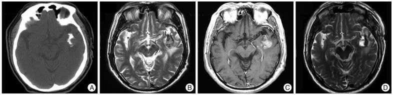
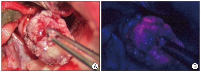
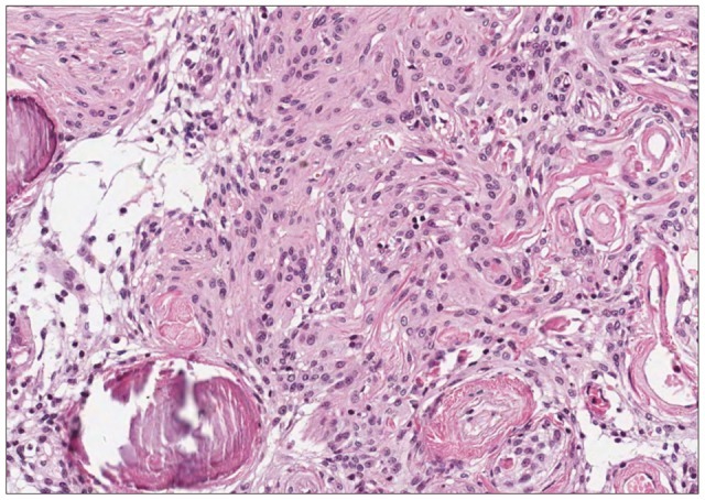
The 5-aminolevulinic acid (5-ALA)-induced tumor fluorescence is a useful intraoperative marker for the diagnosis and the detection of various malignancies, but its use in meningioma is only reported infrequently. In meningioma, a complete resection of the tumor mass is crucial for the prevention of recurrence and postoperative morbidities. Deep sylvian meningioma is a rare type of meningioma where complete tumor removal is complicated by its deep anatomical location and close involvement with the middle cerebral artery. From our experience, 5-ALA-mediated fluorescence facilitated a safe excision whilst preserving critical neurovascular structures. To our best knowledge, this is first report from use of 5-ALA in a deep sylvian meningioma.
Keywords: 5-aminolevulinic acid; Deep sylvian meningioma; Meningioma without dural attachment; Resection.
Figures
Similar articles
-
Deep Sylvian Meningioma without Dural Attachment - A Case Report.NMC Case Rep J. 2019 Mar 21;6(2):51-55. doi: 10.2176/nmccrj.cr.2018-0215. eCollection 2019 Apr. NMC Case Rep J. 2019. PMID: 31016101 Free PMC article.
-
[Deep sylvian meningioma: a case report of a child].No Shinkei Geka. 1994 Dec;22(12):1147-51. No Shinkei Geka. 1994. PMID: 7845511 Review. Japanese.
-
5-Aminolevulinic acid fluorescence-guided surgery for spinal meningioma.World Neurosurg. 2013 Jul-Aug;80(1-2):223.e1-3. doi: 10.1016/j.wneu.2012.12.017. Epub 2012 Dec 13. World Neurosurg. 2013. PMID: 23247024
-
[Deep sylvian meningioma without dural attachment: a case report].No Shinkei Geka. 2011 Nov;39(11):1067-72. No Shinkei Geka. 2011. PMID: 22036818 Review. Japanese.
-
The Utility of 5-Aminolevulinic Acid for Microsurgical Resection of Meningiomas.World Neurosurg. 2020 Jul;139:343. doi: 10.1016/j.wneu.2020.03.178. Epub 2020 Apr 6. World Neurosurg. 2020. PMID: 32272265
Cited by
-
Convexity meningiomas enhanced by sodium fluorescein.Surg Neurol Int. 2014 Jan 14;5:3. doi: 10.4103/2152-7806.124978. eCollection 2014. Surg Neurol Int. 2014. PMID: 24575318 Free PMC article.
-
A huge cerebral parenchymal meningioma in sylvian fissure: case report and literature review.BMC Neurol. 2025 Apr 5;25(1):140. doi: 10.1186/s12883-025-04151-2. BMC Neurol. 2025. PMID: 40188049 Free PMC article. Review.
-
Experience Profiling of Fluorescence-Guided Surgery II: Non-Glioma Pathologies.Brain Tumor Res Treat. 2019 Oct;7(2):105-111. doi: 10.14791/btrt.2019.7.e39. Brain Tumor Res Treat. 2019. PMID: 31686441 Free PMC article.
-
Sylvian Fissure Meningiomas: Case Report and Literature Review.Front Oncol. 2020 Apr 16;10:427. doi: 10.3389/fonc.2020.00427. eCollection 2020. Front Oncol. 2020. PMID: 32373509 Free PMC article.
-
Deep Sylvian Meningioma without Dural Attachment - A Case Report.NMC Case Rep J. 2019 Mar 21;6(2):51-55. doi: 10.2176/nmccrj.cr.2018-0215. eCollection 2019 Apr. NMC Case Rep J. 2019. PMID: 31016101 Free PMC article.
References
-
- Arai T, Tani S, Isoshima A, Nagashima H, Joki T, Takahashi-Fujigasaki J, et al. [Intraoperative photodynamic diagnosis for spinal ependymoma using 5-aminolevulinic acid : technical note] No Shinkei Geka. 2006;34:811–817. - PubMed
-
- Cecchi PC, Campello M, Rizzo P, Mair K, Schwarz A. Atypical meningioma of the sylvian fissure. J Clin Neurosci. 2009;16:1234–1239. - PubMed
-
- Chiocca EA, Boviatsis EJ, Westmark RM, Short MP, Richardson EP, Zervas NT. Deep sylvian fissure meningioma without dural attachment in an adult : case report. Neurosurgery. 1994;35:944–946. discussion 946. - PubMed
-
- Coluccia D, Fandino J, Fujioka M, Cordovi S, Muroi C, Landolt H. Intraoperative 5-aminolevulinic-acid-induced fluorescence in meningiomas. Acta Neurochir (Wien) 2010;152:1711–1719. - PubMed
-
- Grant WE, Hopper C, MacRobert AJ, Speight PM, Bown SG. Photodynamic therapy of oral cancer : photosensitisation with systemic aminolaevulinic acid. Lancet. 1993;342:147–148. - PubMed
Publication types
LinkOut - more resources
Full Text Sources
