

Rabbit maxillary sinus augmentation model with simultaneous implant placement: differential responses to the graft materials
- PMID: 23346463
- PMCID: PMC3543935
- DOI: 10.5051/jpis.2012.42.6.204
Rabbit maxillary sinus augmentation model with simultaneous implant placement: differential responses to the graft materials
Abstract
Purpose: This study was performed to establish an experimental rabbit model for single-stage maxillary sinus augmentation with simultaneous implant placement.
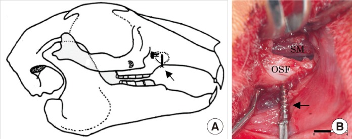
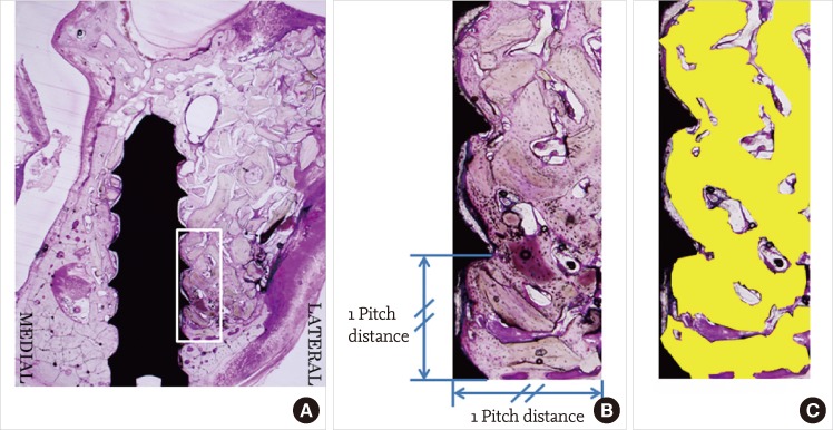
Methods: Twelve mature New Zealand white rabbits were used for the experiments. The rabbit maxillary sinuses were divided into 3 groups according to sinus augmentation materials: blood clot (BC), autogenous bone (AB), and bovine-derived hydroxyapatite (BHA). Small titanium implants were simultaneously placed in the animals during the sinus augmentation procedure. The rabbits were sacrificed 4 and 8 weeks after surgery and were observed histologically. Histomorphometric analyses using image analysis software were also performed to evaluate the parameters related to bone regeneration and implant-bone integration.
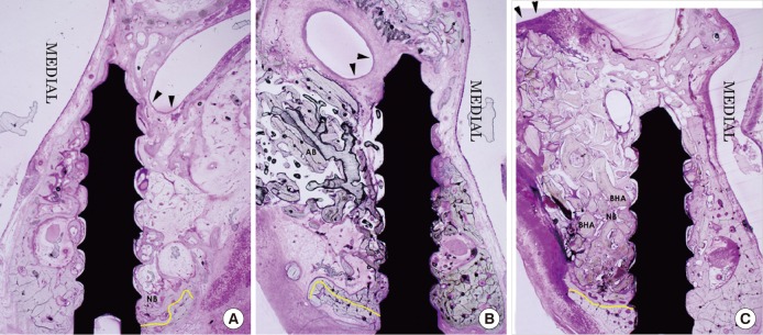
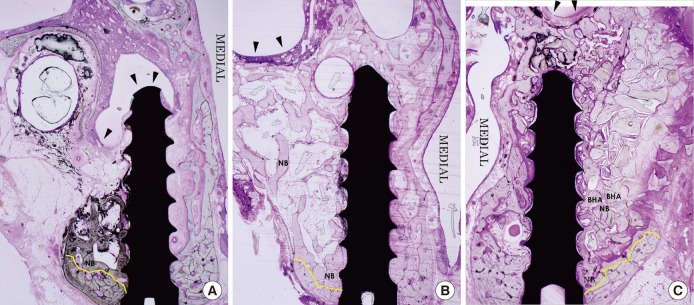
Results: The BC group showed an evident collapse of the sinus membrane and limited new bone formation around the original sinus floor at 4 and 8 weeks. In the AB group, the sinus membrane was well retained above the implant apex, and new bone formation was significant at both examination periods. The BHA group also showed retention of the elevated sinus membrane above the screw apex and evident new bone formation at both points in time. The total area of the mineral component (TMA) in the area of interest and the bone-to-implant contact did not show any significant differences among all the groups. In the AB group, the TMA had significantly decreased from 4 to 8 weeks.
Conclusions: Within the limits of this study, the rabbit sinus model showed satisfactory results in the comparison of different grafting conditions in single-stage sinus floor elevation with simultaneous implant placement. We found that the rabbit model was useful for maxillary sinus augmentation with simultaneous implant placement.
Keywords: Animal models; Bone substitutes; Dental implants; Guided tissue regeneration; Sinus floor augmentation.
Conflict of interest statement
No potential conflict of interest relevant to this article was reported.
Figures
References
-
- Tatum H., Jr Maxillary and sinus implant reconstructions. Dent Clin North Am. 1986;30:207–229. - PubMed
-
- Summers RB. A new concept in maxillary implant surgery: the osteotome technique. Compendium. 1994;15:152, 154–156, 158. - PubMed
-
- Rosen PS, Summers R, Mellado JR, Salkin LM, Shanaman RH, Marks MH, et al. The bone-added osteotome sinus floor elevation technique: multicenter retrospective report of consecutively treated patients. Int J Oral Maxillofac Implants. 1999;14:853–858. - PubMed
-
- Fugazzotto PA. The modified trephine/osteotome sinus augmentation technique: technical considerations and discussion of indications. Implant Dent. 2001;10:259–264. - PubMed
-
- Chen L, Cha J. An 8-year retrospective study: 1,100 patients receiving 1,557 implants using the minimally invasive hydraulic sinus condensing technique. J Periodontol. 2005;76:482–491. - PubMed
LinkOut - more resources
Full Text Sources
Miscellaneous
