Spontaneous complete occlusion of middle cerebral artery aneurysm: case report
- PMID: 23346548
- PMCID: PMC3543918
- DOI: 10.7461/jcen.2012.14.4.309
Spontaneous complete occlusion of middle cerebral artery aneurysm: case report
Abstract
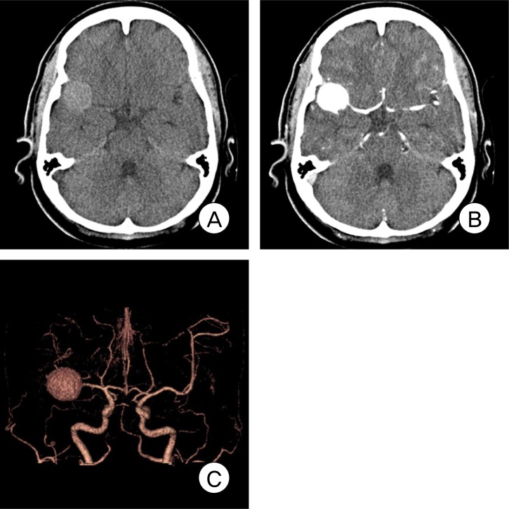
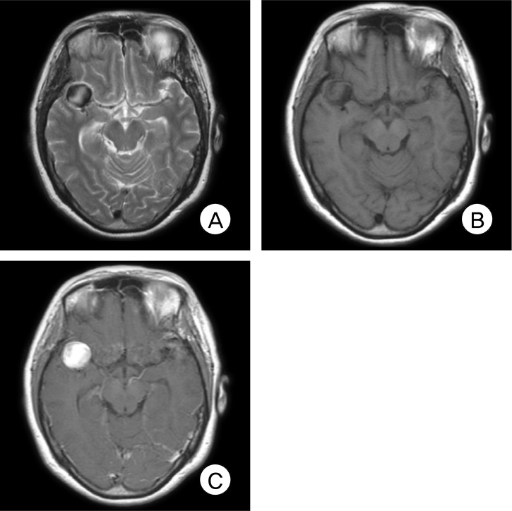
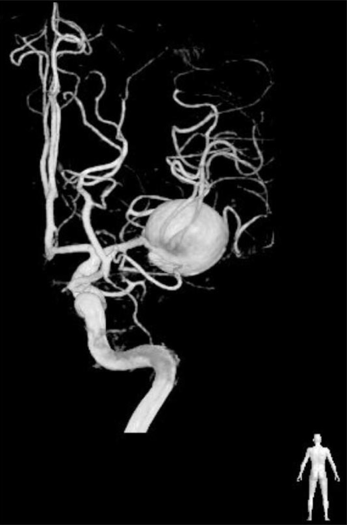
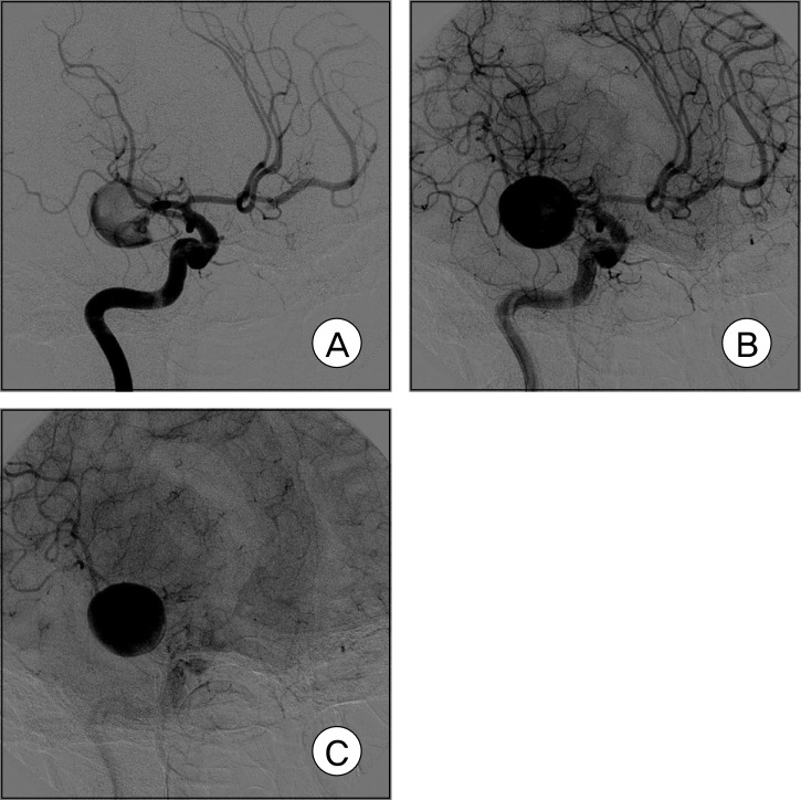
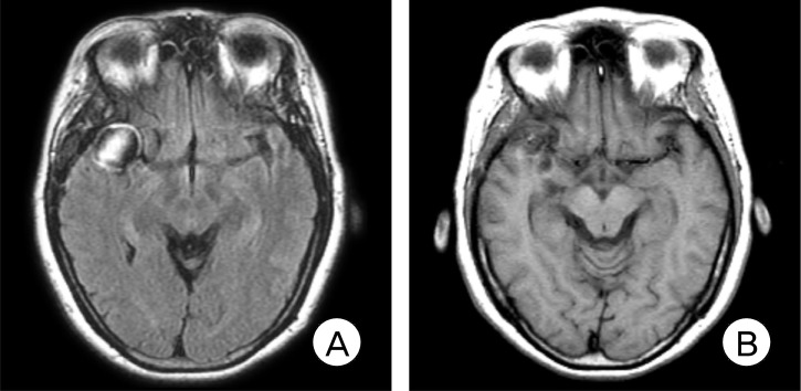
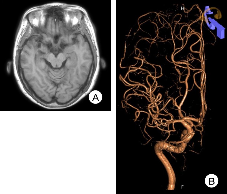
There are few observation papers regarding the natural history of an aneurysm. We report on a case of a completely occluded middle cerebral artery (MCA) aneurysm. A 47-year-old female patient presented with a headache and was diagnosed with rupture of a right MCA aneurysm. Due to a high risk of direct neck clipping, she received conservative treatment after craniotomy and wrapping of her aneurysm. The patient's condition showed improvement, with complete occlusion of the aneurysm and considerable reduction of the aneurysm in size after approximately three years. This is a rare case of an aneurysm of MCA that showed spontaneous resolution. Finally, on the angiogram, characteristics of an aneurysm to occlude spontaneously will be presumed based on literature reviews.
Keywords: Angiography; Middle cerebral artery aneurysm; Spontaneous remission.
Figures
Similar articles
-
[A case of spontaneous middle cerebral artery occlusion associated with a cerebral aneurysm angiographically disappearing after STA-MCA anastomosis].No Shinkei Geka. 1997 Aug;25(8):727-32. No Shinkei Geka. 1997. PMID: 9266566 Japanese.
-
[Posterior cerebral aneurysm associated with complete occlusion of the middle cerebral artery caused subarachnoid hemorrhage: a case report].No Shinkei Geka. 1996 Dec;24(12):1107-11. No Shinkei Geka. 1996. PMID: 8974093 Review. Japanese.
-
Coil embolization for intracranial aneurysms: an evidence-based analysis.Ont Health Technol Assess Ser. 2006;6(1):1-114. Epub 2006 Jan 1. Ont Health Technol Assess Ser. 2006. PMID: 23074479 Free PMC article.
-
Disappearance of Ruptured Posterior Cerebral Artery Aneurysm Associated with Internal Carotid Artery Occlusion After Superficial Temporal Artery-to-Middle Cerebral Artery Bypass.World Neurosurg. 2018 Aug;116:178-181. doi: 10.1016/j.wneu.2018.05.096. Epub 2018 May 23. World Neurosurg. 2018. PMID: 29803057
-
Completely Thrombosed Distal Middle Cerebral Artery Aneurysm Mimicking a Cavernous Angioma: Case Report and Review of the Literature.World Neurosurg. 2017 Jul;103:955.e1-955.e4. doi: 10.1016/j.wneu.2017.04.172. Epub 2017 May 9. World Neurosurg. 2017. PMID: 28499904 Review.
Cited by
-
Clipping first policy for middle cerebral artery aneurysm: A single-center cohort study.Surg Neurol Int. 2024 Dec 27;15:474. doi: 10.25259/SNI_756_2024. eCollection 2024. Surg Neurol Int. 2024. PMID: 39777182 Free PMC article.
-
Thrombosis and recanalization of small saccular cerebral aneurysm : two case reports and a suggestion for possible mechanism.J Korean Neurosurg Soc. 2014 May;55(5):280-3. doi: 10.3340/jkns.2014.55.5.280. Epub 2014 May 31. J Korean Neurosurg Soc. 2014. PMID: 25132936 Free PMC article.
References
-
- International Study of Unruptured Intracranial Aneurysms Investigators. Unruptured intracranial aneurysms-risk of rupture and risks of surgical intervention. N Engl J Med. 1998 Dec;339(24):1725–1733. - PubMed
-
- De Lange SA. [A case of spontaneous thrombosis of a cerebral arteriovenous aneurysm] Ned Tijdschr Geneeskd. 1955 Mar;99(13):944–946. Dutch. - PubMed
-
- Vasconcellos LP, Flores JAC, Conti MLM, Veiga JCE, Lancellotti CLP. Spontaneous thrombosis of internal carotid artery: a natural history of giant carotid cavernous aneurysms. Arq Neuropsiquiatr. 2009 Jun;67(2A):278–283. - PubMed
-
- Deshmukh VR, Kakarla UK, Figueiredo EG, Zabramski JM, Spetzler RF. Long-term clinical and angiographic follow-up of unclippable wrapped intracranial aneurysms. Neurosurgery. 2006 Mar;58(3):434–442. discussion 434-42. - PubMed
-
- Fujiwara S, Fujii K, Nishio S, Fukui M. Long-term results of wrapping of intracranial ruptured aneurysms. Acta Neurochir (Wien) 1990;103(1-2):27–29. - PubMed