. 2013 Jan 22:3:2.
doi: 10.3389/fonc.2013.00002. eCollection 2013.

Histopathology of gastrointestinal neuroendocrine neoplasms

Affiliations

Histopathology of gastrointestinal neuroendocrine neoplasms

Kenichi Hirabayashi et al. Front Oncol. .

Abstract

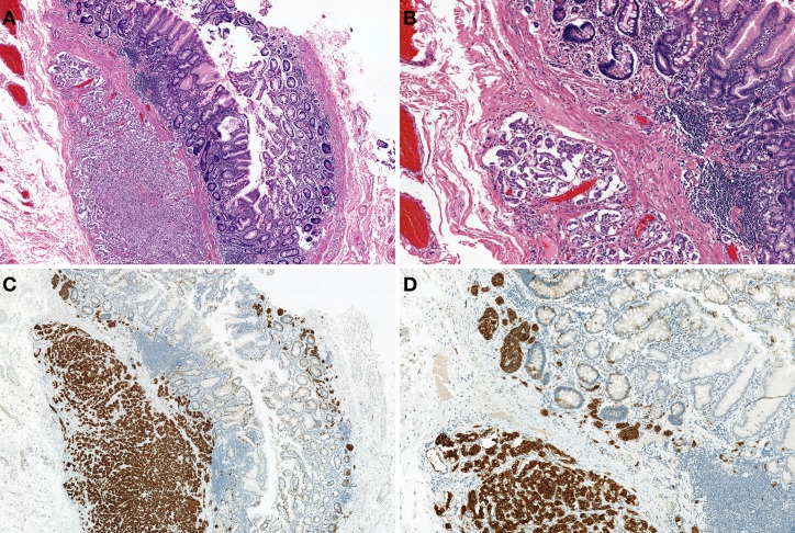
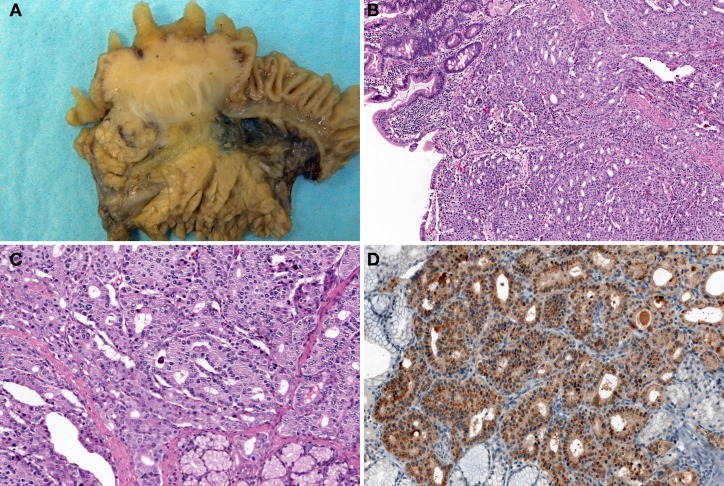
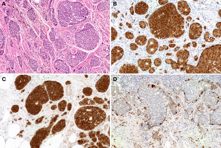
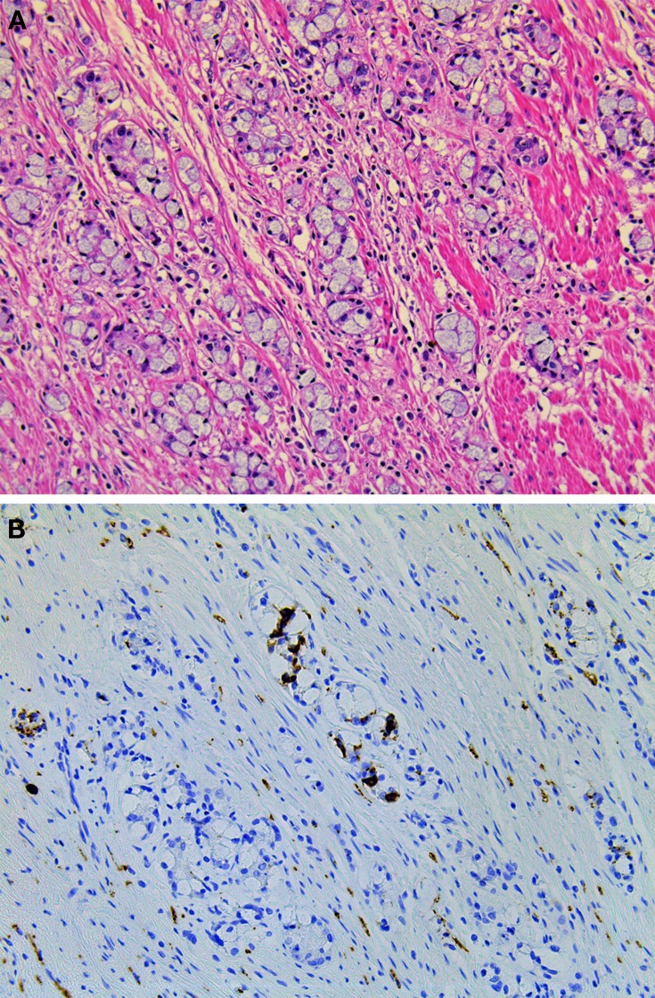
Gastrointestinal neuroendocrine neoplasms (GI-NENs) arise from neuroendocrine cells distributed mainly in the mucosa and submucosa of the gastrointestinal tract. In 2010, the World Health Organization (WHO) classification of NENs of the digestive system was changed, categorizing these tumors as grade 1 neuroendocrine tumor (NET), grade-2NET, neuroendocrine carcinoma (large- or small-cell type), or mixed adenoneuroendocrine carcinoma (MANEC). Such a classification is based on the Ki-67 index and mitotic count in histological material. For the accurate pathological diagnosis and grading of NENs, it is important to clearly recognize the characteristic histological features of GI-NENs and to understand the correct method of counting Ki-67 and mitoses. In this review, we focus on the histopathological features of GI-NENs, particularly regarding biopsy and cytological diagnoses, neuroendocrine markers, genetic and molecular features, and the evaluation of the Ki-67 index and mitotic count. In addition, we will address the histological features of GI-NEN in specific organs.

Keywords: carcinoid; gastrointestinal tract; neuroendocrine carcinoma; neuroendocrine marker; neuroendocrine neoplasm.


Figures

Figure 1
Figure 1
Rectal neuroendocrine tumor (NET): a solitary sessile mass with slight ulceration.
Figure 2
Figure 2
Neuroendocrine carcinoma (NEC) at the esophagogastric junction: a large ulcerated mass.
Figure 3
Figure 3
“Crush”artifacts of esophageal small cell carcinoma (upper right) [hematoxylinand eosin (H and E) stain].
Figure 4
Figure 4
Combined tumors of neuroendocrine carcinoma (NEC) (left) and squamous cell carcinoma (right) (H and E stain).
Figure 5
Figure 5
Type-1 neuroendocrine neoplasm (NEN) of the stomach. Tumor cells proliferate in a trabecular fashion in the submucosa. Enterochromaffin-like-cell (ECL-cell) hyperplasia is scattered in the mucosa (H and E stain, A: low power view, B: high power view). Synaptophysin is positive for tumor and ECL-cell hyperplasia (C: low power view, D: high power view).
Figure 6
Figure 6
Gastrin-producing NETs of the duodenum. Tumor shows trabecular and pseudoglandular patterns (H and E stain, A: low power view, B: high power view) with immunohistochemical gastrin expression (C). Gastrin-producing NETs metastasize to the lymph node (D).
Figure 7
Figure 7
Somatostatin-producing NETs arising from the papilla of Vater. Macroscopic findings show the solid, polypoid mass in the papilla of Vater (A). The tumor is arranged in a trabecular pattern with pseudoglandular structures (H and E stain, B: low power view, C: high power view). Psammoma bodies are present (C). Immunohistochemically, the tumor cells are positive for somatostatin (D).
Figure 8
Figure 8
EC-cell NETs of the appendix. The tumor proliferates in an insular growth pattern, with solid to cribriform tumor structures (H and E stain, A). The tumor cells are positive for synaptophysin (B) and serotonin (C). S-100 protein positivity is observed in sustentacular-like cells (D).
Figure 9
Figure 9
Goblet cell carcinoid of the appendix. (A) Tumor cells of goblet cell carcinomas possess intracytoplasmic mucus with small, compressed nuclei. (B) Tumor cells are focally positive for synaptophysin.
Figure 10
Figure 10
Microscopic findings of rectal NET. Rectal NET shows a trabecular proliferating pattern (A: low power view, B: high power view). Immunohistochemically, tumor cells of rectal NET are positive for synaptophysin (C) but negative for chromogranin A (D).

Similar articles

Cited by

References

    1. Acs G., McGrath C. M., Gupta P. K. (2000). Duodenal carcinoid tumor: report of a case diagnosed by endoscopic ultrasound-guided fine-needle aspiration biopsy with immunocytochemical correlation. Diagn. Cytopathol. 23, 183–186 10.1002/1097-0339(200009)23:3<183::AID-DC8>3.0.CO;2-4 - DOI - PubMed
    1. Al-Khafaji B., Noffsinger A. E., Miller M. A., Devoe G., Stemmermann G. N., Fenoglio-Preiser C. (1998). Immunohistologic analysis of gastrointestinal and pulmonary carcinoid tumors. Hum. Pathol. 29, 992–999 - PubMed
    1. Alsaad K. O., Serra S., Schmitt A., Perren A., Chetty R. (2007). Cytokeratins 7 and 20 immunoexpression profile in goblet cell and classical carcinoids of appendix. Endocr. Pathol. 18, 16–22 - PubMed
    1. Anlauf M., Perren A., Meyer C. L., Schmid S., Saremaslani P., Kruse M. L., et al. (2005). Precursor lesions in patients with multiple endocrine neoplasia type 1-associated duodenal gastrinomas. Gastroenterology 128, 1187–1198 - PubMed
    1. Arnason T., Sapp H. L., Barnes P. J., Drewniak M., Abdolell M., Rayson D. (2011a). Immunohistochemical expression and prognostic value of ER, PR and HER2/neu in pancreatic and small intestinal neuroendocrine tumors. Neuroendocrinology 93, 249–258 10.1159/000326820 - DOI - PubMed

LinkOut - more resources