

Early knee changes in dancers identified by ultra-high-field 7 T MRI
- PMID: 23346987
- PMCID: PMC3723761
- DOI: 10.1111/sms.12039
Early knee changes in dancers identified by ultra-high-field 7 T MRI
Abstract
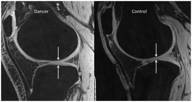
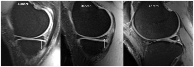
We aimed to determine whether a unique, ultra-high-field 7 T magnetic resonance imaging (MRI) scanner could detect occult cartilage and meniscal injuries in asymptomatic female dancers. This study had Institutional Review Board approval. We recruited eight pre-professional female dancers and nine non-athletic, female controls. We scanned the dominant knee on a 7 T MRI scanner using a three-dimensional fast low-angle shot sequence and a proton density, fast spin-echo sequence to evaluate cartilage and menisci, respectively. Two radiologists scored cartilage (International Cartilage Repair Society classification) and meniscal (Stoller classification) lesions. We applied two-tailed z- and t-tests to determine statistical significance. There were no cartilage lesions in dancers or controls. For the medial meniscus, the dancers demonstrated higher mean MRI score (2.38 ± 0.61 vs 1.0 ± 0.97, P < 0.0001) and higher frequency of mean grade 2 lesions (88% vs 11%, P < 0.01) compared with the controls. For the lateral meniscus, there was no difference in score (0.5 ± 0.81 vs 0.5 ± 0.78, P = 0.78) in dancers compared with the control groups. Asymptomatic dancers demonstrate occult medial meniscal lesions. Because this has been described in early osteoarthritis, close surveillance of dancers' knee symptoms and function with appropriate activity modification may help maintain their long-term knee health.
Keywords: 7 Tesla; MRI; dancers; knee; meniscus.
© 2013 John Wiley & Sons A/S. Published by John Wiley & Sons Ltd.
Figures
References
-
- Banerjee S, Krug R, Carballido-Gamio J, Kelley DA, Xu D, Vigneron DB, Majumdar S. Rapid in vivo musculoskeletal MR with parallel imaging at 7T. Magn Reson Med. 2008;59:655–660. - PubMed
-
- Brittberg M, Winalski CS. Evaluation of cartilage injuries and repair. J Bone Joint Surg Am. 2003;85-A(Suppl 2):58–69. - PubMed
Publication types
MeSH terms
Grants and funding
LinkOut - more resources
Full Text Sources
Other Literature Sources
Medical
