Review
. 2013 Mar;68(3):274-84.
doi: 10.1111/all.12103. Epub 2013 Jan 25.

Eosinophil-derived cytokines in health and disease: unraveling novel mechanisms of selective secretion

Affiliations
Review

Eosinophil-derived cytokines in health and disease: unraveling novel mechanisms of selective secretion

R C N Melo et al. Allergy. 2013 Mar.

Abstract

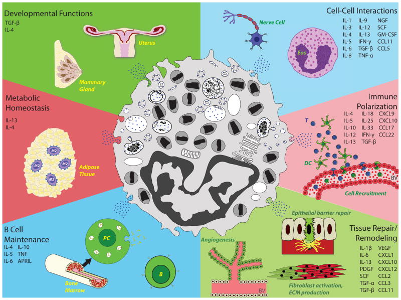
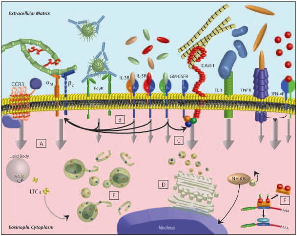
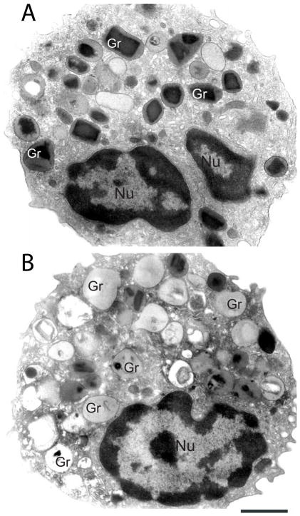
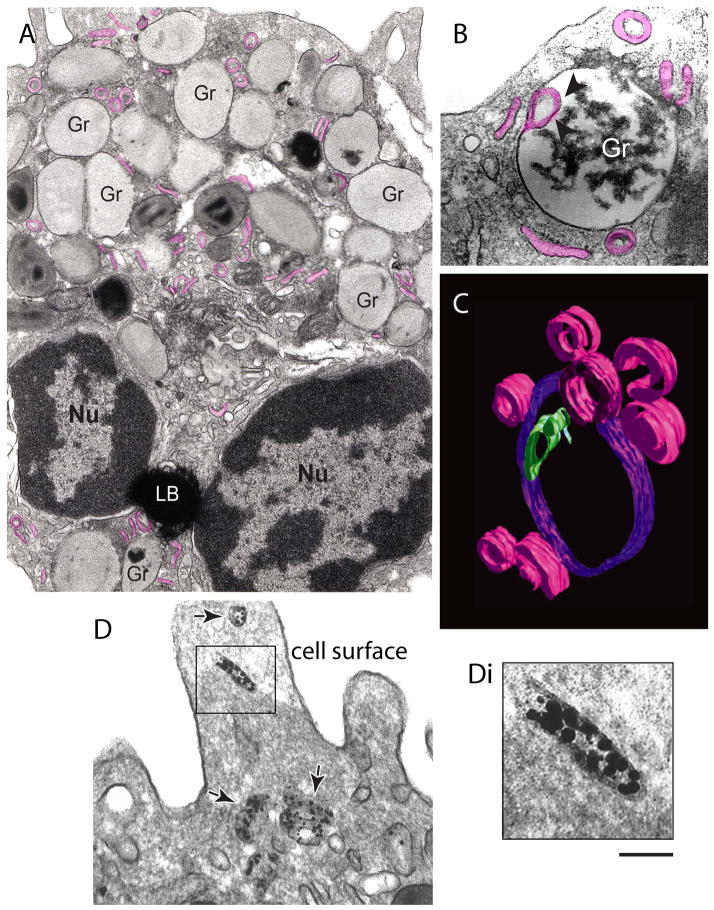
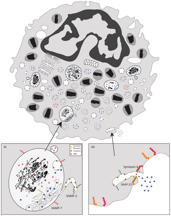
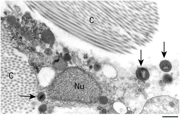
Over the past two decades, our understanding of eosinophils has evolved from that of categorically destructive effector cells to include active participation in immune modulation, tissue repair processes, and normal organ development, in both health and disease. At the core of their newly appreciated functions is the capacity of eosinophils to synthesize, store within intracellular granules, and very rapidly secrete a highly diverse repertoire of cytokines. Mechanisms governing the selective secretion of preformed cytokines from eosinophils are attractive therapeutic targets and may well be more broadly applicable to other immune cells. Here, we discuss recent advances in deciphering pathways of cytokine secretion, both from intact eosinophils and from tissue-deposited cell-free eosinophil granules, extruded from eosinophils undergoing a lytic cell death.


Conflict of interest statement

Conflict of interest

The authors declare no conflict of interest.

Figures

Figure 1
Figure 1. Biological effects of eosinophil-derived cytokines in health and disease
Eosinophils secrete numerous cytokines with varied biological functions in health (left panels) and disease (right panels). Shown is an abridged list of eosinophil-derived cytokines compiled from(6, 89, 90). M2=alternatively activated macrophage; PC=plasma cell; B=B cell; BV=blood vessel; Eos=eosinophil; T=T cell; DC=dendritic cell.
Figure 2
Figure 2. Stimulus-induced activation of intracellular cytokine secretory pathways in eosinophils
Eosinophil cytokine secretion is initiated through interactions of a wide variety of eosinophil secretagogues, including soluble factors and tissue components, with their respective receptors. Intracellular signal transduction pathways (vertical gray arrows) are initiated downstream of single receptor activation (narrow gray arrows), or following the synergistic activation of multiple receptors (wide gray arrows). Stimuli may elicit intracellular signaling intermediaries (A), or promote expression and/or activation of eosinophil-expressed adhesion molecules (B, C), in order to accomplish cytokine secretion. Secreted cytokines are either synthesized de novo and shuttled through golgi apparatus for packaging and secretion (D), translated from nascent pools of cytoplasmic mRNA (E), or sorted and mobilized from preformed intracellular granule caches into secretory vesicles for delivery to cell surface (F).
Figure 3
Figure 3. Ultrastructural images of human eosinophils showing normal appearance (A) and piecemeal degranulation (PMD) characteristics (B)
In (A), the cytoplasm is packed with specific (secretory) granules (Gr) containing an internal often electron-dense crystalline core surrounded by an electron-lucent matrix. (B) After stimulation with physiological stimuli, granules (Gr) exhibit progressive emptying of their contents classically associated with PMD. Disassembled cores and reduced electron-density are frequently observed. Cells were stimulated with eotaxin as described (54) and prepared for conventional transmission electron microscopy. Nu, nucleus. Bar, 900 nm.
Figure 4
Figure 4. Morphology of human stimulated eosinophils undergoing piecemeal degranulation (PMD)
(A) Enlarged, emptying secretory granules (Gr) and large tubular carriers termed eosinophil sombrero vesicles (EoSVs, highlighted in pink) are observed in the cytoplasm. In (B), a highly mobilized granule (Gr) with intragranular membranes (arrowheads) is seen in high magnification. Note different profiles of EoSVs surrounding or in contact with the granule. In (C), a three-dimensional (3D) model based on electron tomographic slices show the structural organization of EoSVs (pink). The granule limiting membrane is highlighted in blue and intragranular membranes in green. (D) Immunonanogold electron microscopy (EM) for major basic protein (MBP) reveals labeling at EoSVs lumina (arrows). Cells were stimulated with eotaxin (A, C, D) or platelet-activating factor (PAF) (B) as before (54) and prepared for conventional (A, B) or immunanogold (C) EM. The 3D model was constructed from serial virtual slices extracted from reconstructions of a human eosinophil analyzed by fully automated electron tomography at 200 kV. Figure 4C was reprinted from (91) with permission. Nu, nucleus; LB, lipid body. Bar, 400 nm (A), 200 nm (B, C), 500 nm (D).
Figure 5
Figure 5. Proposed model of receptor-mediated chaperoning of cognate cytokines from intragranular stores
Cytokine receptor chains, expressed within granules and mobilized downstream of cell stimulation, sequester and chaperone cognate cytokine ligands into emerging secretory vesicles (Ai). Unlike MBP, which exhibits a free luminal pattern of expression within secretory vesicles (red circles), granule-derived cytokines (blue circles) remain bound to receptor chaperones within secretory vesicles. Docking and fusion of secretory vesicles to plasma membrane is directed through complexes formed between vesicle-expressed VAMP-2 and plasma membrane-expressed Syntaxin-4 and SNAP-23 (Aii).
Figure 6
Figure 6. Ultrastructure of a tissue human eosinophil undergoing cytolysis
Note the disintegrating nucleus (Nu) and the entire membrane-bound secretory granules (arrows) in the surrounding tissue. Tissue eosinophils were present in a skin biopsy performed on a patient with breast cancer who underwent treatment using rhSCF (recombinant human Stem Cell Factor). CF, collagen fibrils. Bar, 800 nm.

Similar articles

Cited by

References

    1. Voehringer D, Shinkai K, Locksley RM. Type 2 immunity reflects orchestrated recruitment of cells committed to IL-4 production. Immunity. 2004;20(3):267–77. - PubMed
    1. Sabin EA, Kopf MA, Pearce EJ. Schistosoma mansoni egg-induced early IL-4 production is dependent upon IL-5 and eosinophils. J Exp Med. 1996;184(5):1871–8. - PMC - PubMed
    1. Voehringer D, Reese TA, Huang X, Shinkai K, Locksley RM. Type 2 immunity is controlled by IL-4/IL-13 expression in hematopoietic non-eosinophil cells of the innate immune system. J Exp Med. 2006;203(6):1435–46. - PMC - PubMed
    1. Jacobsen EA, Ochkur SI, Pero RS, Taranova AG, Protheroe CA, Colbert DC, et al. Allergic pulmonary inflammation in mice is dependent on eosinophil-induced recruitment of effector T cells. J Exp Med. 2008;205(3):699–710. - PMC - PubMed
    1. Walsh ER, Sahu N, Kearley J, Benjamin E, Kang BH, Humbles A, et al. Strain-specific requirement for eosinophils in the recruitment of T cells to the lung during the development of allergic asthma. J Exp Med. 2008;205(6):1285–92. - PMC - PubMed

Publication types