Review
. 2013 Apr;10(4):499-509.
doi: 10.1517/17425247.2013.764864. Epub 2013 Jan 25.

Hydrogels for lentiviral gene delivery

Affiliations
Review

Hydrogels for lentiviral gene delivery

Stephanie K Seidlits et al. Expert Opin Drug Deliv. 2013 Apr.

Abstract

Introduction: Gene delivery from hydrogel biomaterials provides a fundamental tool for a variety of clinical applications including regenerative medicine, gene therapy for inherited disorders and drug delivery. The high water content and mild gelation conditions of hydrogels support their use for gene delivery by preserving activity of lentiviral vectors and acting to shield vectors from any host immune response.

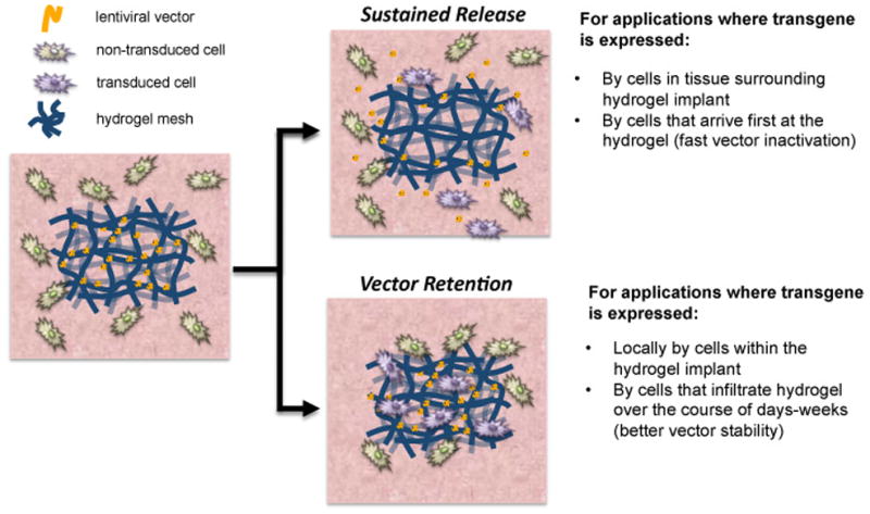
Areas covered: Strategies to control lentiviral entrapment within and retention/release from hydrogels are reviewed. The authors discuss the ability of hydrogel design parameters to control the transgene expression profile and the capacity of hydrogels to protect vectors from (and even modulate) the host immune response.

Expert opinion: Delivery of genetic vectors from scaffolds provides a unique opportunity to capitalize on the potential synergy between the biomaterial design for cell processes and gene delivery. Hydrogel properties can be tuned to directly control the events that determine the tissue response to controlled gene delivery, which include the extent of cell infiltration, preservation of vector activity and vector retention. While some design parameters have been identified, numerous opportunities for investigation are available in order to develop a complete model relating the biomaterial properties and host response to gene delivery.


Conflict of interest statement

Declaration of Interests

The authors thank the NIH (R01 EB005678, R01 EB003806, R21 EB006520 and PL1 EB008542, a P30 Biomaterials Core within the Oncofertility Consortium Roadmap Grant) for support.

Figures

Figure 1
Figure 1
Gene delivery strategies using hydrogels. The hydrogel design parameters for delivery of gene therapy vectors can be modulated to achieve different transgene expression profiles. Hydrogels can be designed to enable sustained release of vectors to target the cells surrounding the hydrogel. Alternatively, hydrogels can be designed to retain vectors within the scaffold to target infiltrating cells and better preserve vector activity.
Figure 2
Figure 2
In vivo transgene expression from lentivirus loaded hydrogels with and without macroporous structure. (a) Representative bioluminescence images following subcutaneous implantation of hydrogels. Three virus loading configurations into the PEG hydrogels are shown: (i) non-macroporous PEG (no gelatin) (PEGnp) hydrated with virus solution following gelation, (ii) PEG encapsulating gelatin microspheres (PEGmp) hydrated with virus solution following gelation, and (iii) PEG encapsulating gelatin microspheres that have been loaded with virus prior to gelation (pPEGmp) by swelling gelatin microspheres with virus solution. A gelatin only control is also shown. (b) Transgene expression was measured as integrated photon flux (photons/s) using an IVIS bioluminescence imaging system (b). Asterisks represent statistical difference (p < 0.05) relative to the PEGmp condition at each time point based on a KruskaleWallis test. (c), (d) Hematoxylin and eosin staining showing cell infiltration into PEGmp (c) and PEGnp (d) hydrogels 6 wks after subcutaneous implantation. The labels P and T denote PEG and tissue surrounding the implant, respectively. White lines denote hydrogel-tissue interface. Scale bars = 100 μm. Reprinted from Biomaterials, 33, Shepard JA, Virani FR, Goodman AG, Gossett TD, Shin S, Shea LD, Hydrogel macroporosity and the prolongation of transgene expression and the enhancement of angiogenesis, 7412–7421, 2012, with permission from Elsevier.
Figure 3
Figure 3
In vivo transgene expression from hydrogel-filled polylactide-glycolide scaffolds as a function of time. (a) Hydrogel (collagen, fibrin, or alginate) precursors were mixed with a lentivirus encoding for the reporter gene luciferase, loaded into the pores of a PLG scaffold, and implanted in the fat pad of mice. Transgene expression was then measured using an in vivo bioluminesence imaging. Letters indicate statistically significant differences (p < 0.05) compared to (a) alginate or (c) collagen analyzed by a Kruskal–Wallis test (n= 3). (b) Representative bioluminescence images from scaffolds at 3 and 14 days post-implantation. Reprinted with permission from Springer Science+Business Media: Drug Del Transl Res, Hydrogels to modulate lentivirus delivery in vivo from microporous tissue engineering scaffolds, 1, 2011, 91–101, Aviles MO, Shea LD, Figure 4.
Figure 4
Figure 4
In vivo transgene expression within fibrin hydrogels. Fibrin hydrogels containing either virus alone or HA/virus complexes were transplanted subcutaneously and expression profile was monitored for 28 days. (a) Represenative images of in vivo luciferase expression using bioluminescence imaging. (b) Quantification of luminescence as a function of time (n=6). Values at day 21 are statsitically different. Reprinted from J Control Rel, 157, Kidd ME, Shin S, Shea LD, Fibrin hydrogels for lentiviral gene delivery in vitro and in vivo, 80–85, 2012, with permission from Elsevier.

References

    1. Flotte TR, Trapnell BC, Humphries M, et al. Phase 2 clinical trial of a recombinant adeno-associated viral vector expressing alpha1-antitrypsin: Interim results. Human Gene Ther. 2011;22:1239–47. - PMC - PubMed
    1. LeWitt PA, Rezai AR, Leehey MA, et al. AAV2-GAD gene therapy for advanced Parkinson’s disease: A double-blind, sham-surgery controlled, randomised trial. Lancet Neurol. 2011;10:309–19. - PubMed
    1. Nathwani AC, Tuddenham EG, Rangarajan S, et al. Adenovirus-associated virus vector-mediated gene transfer in hemophilia B. New Engl J Med. 2011;365:2357–65. - PMC - PubMed
    1. Bowles DE, McPhee SW, Li C, et al. Phase 1 gene therapy for Duchenne muscular dystrophy using a translational optimized AAV vector. Mol Ther. 2012;20:443–55. - PMC - PubMed
    1. Partridge KA, Oreffo ROC. Gene delivery in bone tissue engineering: Progress and prospects using viral and nonviral strategies. Tissue Eng. 2004;10:295–307. - PubMed

Publication types