Review
. 2013:103:1-33.
doi: 10.1016/B978-0-12-385979-2.00001-0.

Developmental checkpoints and feedback circuits time insect maturation

Affiliations
Review

Developmental checkpoints and feedback circuits time insect maturation

Kim F Rewitz et al. Curr Top Dev Biol. 2013.

Abstract

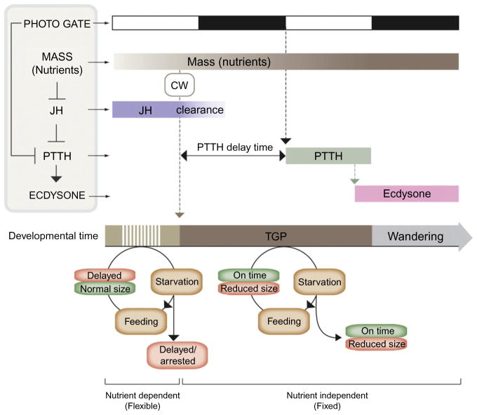
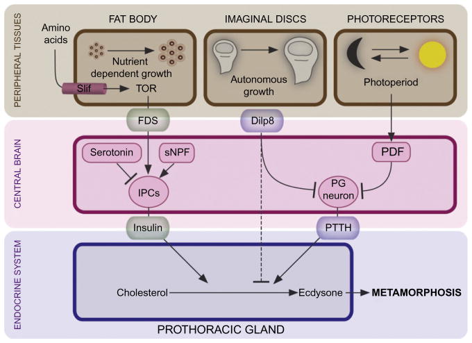
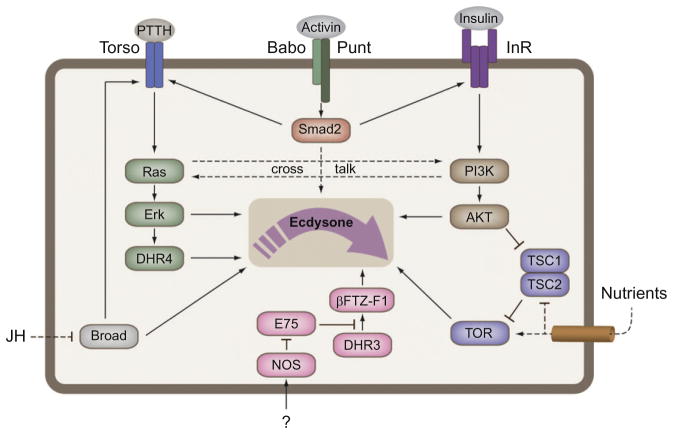
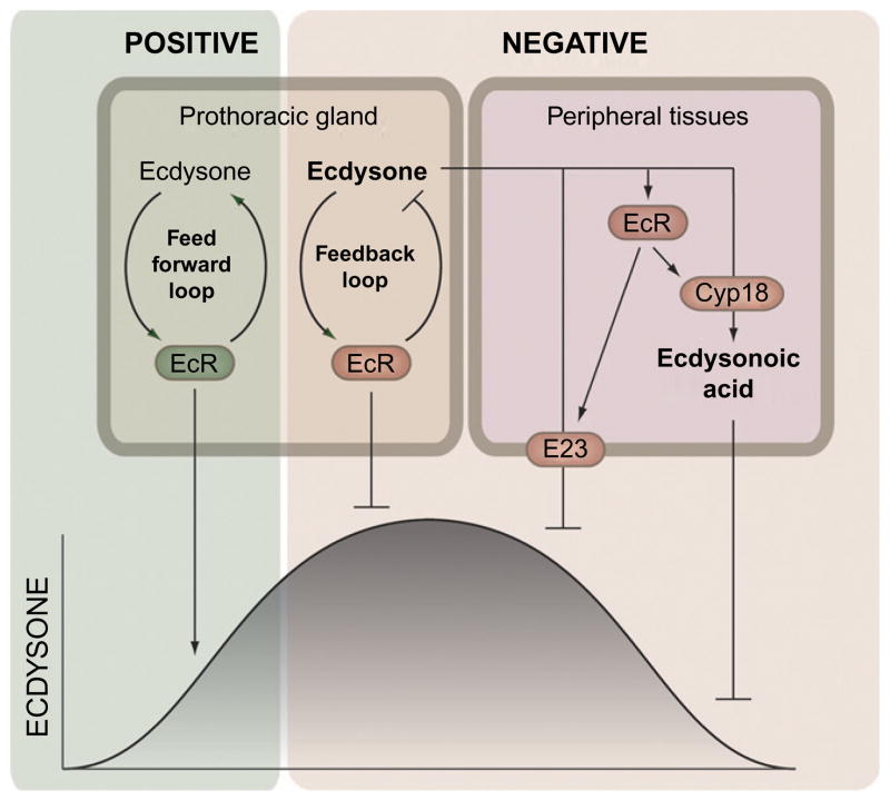
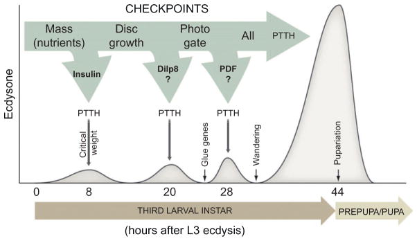
The transition from juvenile to adult is a fundamental process that allows animals to allocate resource toward reproduction after completing a certain amount of growth. In insects, growth to a species-specific target size induces pulses of the steroid hormone ecdysone that triggers metamorphosis and reproductive maturation. The past few years have seen significant progress in understanding the interplay of mechanisms that coordinate timing of ecdysone production and release. These studies show that the neuroendocrine system monitors complex size-related and nutritional signals, as well as external cues, to time production and release of ecdysone. Based on results discussed here, we suggest that developmental progression to adulthood is controlled by checkpoints that regulate the genetic timing program enabling it to adapt to different environmental conditions. These checkpoints utilize a number of signaling pathways to modulate ecdysone production in the prothoracic gland. Release of ecdysone activates an autonomous cascade of both feedforward and feedback signals that determine the duration of the ecdysone pulse at each developmental transitions. Conservation of the genetic mechanisms that coordinate the juvenile-adult transition suggests that insights from the fruit fly Drosophila will provide a framework for future investigation of developmental timing in metazoans.


Figures

Figure 1.1
Figure 1.1
Hormonal regulation of body size and timing of metamorphosis. According to classical work the larval accumulation of mass corresponding to the critical weight results in the breakdown of JH that inhibits PTTH release. Above critical weight, PTTH is release after a delay period determined by the time of JH clearance and the photo-period gating of PTTH. Starvation before critical weight results in a developmental delay of pupariation. If feeding is resumed critical weight is attained and normal body size achieved. In contrast, larvae starved above critical weight, when the metamorphic program is activated, pupariate on time but with a reduced size. As critical weight is independent of nutrition, final size is determined by the amount of growth in the interval between critical weight and cessation of feeding (wandering) called the terminal growth period (TGP). Photo gate, a PTTH gating mechanism imposed by the photo-period; CW, critical weight.
Figure 1.2
Figure 1.2
The developmental timing system monitors environmental (nutrient status and photoperiod) and developmental (disks maturation) cues. The fat body acts as a nutrient sensor that coordinates nutrient uptake with systemic growth and developmental timing. In growth permissive environments, the fat body secretes an unknown fat body-derived signal (FDS), in response to dietary amino acids, that stimulates release of insulin from the insulin producing cells (IPCs) of the brain which acts on the PG and stimulates ecdysone release. Serotonin and short neuropeptide F (sNPF) also impinge on the IPCs and regulate insulin release. In addition to insulin, the developmental timing program checks the status of the imaginal disks. Disk growth and maturation is controlled by a tissue-autonomous program that via Dilp8 crosstalks with the neuroendocrine system. Disks secrete Dilp8 which suppresses ecdysone release presumably by inhibition of PTTH release from the PG neurons until they have completed a certain amount of growth or regenerated from tissue damage. Superimposed on this, the clock neurons producing the pigment dispersing factor (PDF) impinge on the PG neurons and regulate PTTH release according to the photoperiod.
Figure 1.3
Figure 1.3
Mechanisms converging on the PG time ecdysone production. TGFβ/Activin is required for normal expression of the insulin receptor (InR) and torso, which provides glandular competence to PTTH (developmental) and insulin (nutrient) cues. Furthermore, the PG harbors a TOR-dependent nutrients sensor that presumably allows compensation for poor nutrient environments. Under such conditions, ecdysone release is delayed which prolongs the growth period. Broad is required for normal expression of the Halloween genes encoding the enzymes mediating ecdysone synthesis, although it is not clear if JH regulates broad in the PG. Nitric oxide (NO) generated by the nitric oxide synthase (NOS) regulates nuclear receptor signaling in the PG. In turn, NO inhibits E75, a repressor of DHR3 which then activates expression of βFTZ-F1, a nuclear receptor required for expression of at least two key ecdysone biosynthetic enzymes, Phantom and Disembodied.
Figure 1.4
Figure 1.4
Feedback control shapes the ecdysone pulses. A short positive and a long negative feedback loop are believed to operate in the PG to control synthesis of ecdysone. The short positive feedforward loop presumably amplifies the ecdysone signal causing a fast increase of the titer. On the other hand, a long negative feedback loop shuts off the PG which allows peripheral mechanisms to clear ecdysone from the system. Ecdysone induces two feedback mechanisms, in tissues peripheral to the PG, which are eventually responsible for lowering cellular levels (E23) and the decline of the titer (Cyp18). Ecdysone induces Cyp18 that eliminates ecdysone by converting it into the inactive ecdysonoic acid. The ecdysone-inducible E23 encodes an ABC transporter believed to pump ecdysone out of the cells. Together these feedback mechanisms determine the duration of the ecdysone pulses.
Figure 1.5
Figure 1.5
Checkpoint controls of developmental timing of metamorphosis. The figure shows a proposed model for checkpoint control of ecdysone release and timing of pupariation. Accumulation of nutrients corresponding to the critical weight presumably results in the insulin dependent production of the critical weight ecdysone peak 8 h after L3 ecdysis. Maturation and patterning of the imaginal disks and the photoperiod are also verified by the endocrine system before development can proceed. These checkpoints may potentially translate into to the other low-level ecdysone peaks 20 and 28 h after L3 ecdysis. When all checkpoints are cleared, developmental transition to the metamorphic stage can take place.

Similar articles

Cited by

References

    1. Ahmed ML, Ong KK, Dunger DB. Childhood obesity and the timing of puberty. Trends in Endocrinology and Metabolism. 2009;20:237–242. - PubMed
    1. Ainsley JA, Kim MJ, Wegman LJ, Pettus JM, Johnson WA. Sensory mechanisms controlling the timing of larval developmental and behavioral transitions require the Drosophila DEG/ENaC subunit, Pickpocket1. Developmental Biology. 2008;322:46–55. - PubMed
    1. Baehrecke EH. Steroid regulation of programmed cell death during Drosophila development. Cell Death and Differentiation. 2000;7:1057–1062. - PubMed
    1. Beadle G, Tatum E, Glancy C. Food level in relation to rate of development and eye pigmentation in Drosophila melanogaster. The Biological Bulletin. 1938;75:447–462.
    1. Beydon P, Lafont R. Feedback inhibition of ecdysone production by 20-hydroxyecdysone in Pieris brassicae pupae. Journal of Insect Physiology. 1983;29:529–533.

Publication types

LinkOut - more resources