. 2013 Apr;34(11):2641-54.
doi: 10.1016/j.biomaterials.2012.12.048. Epub 2013 Jan 21.

Decellularized musculofascial extracellular matrix for tissue engineering

Affiliations

Decellularized musculofascial extracellular matrix for tissue engineering

Lina Wang et al. Biomaterials. 2013 Apr.

Abstract

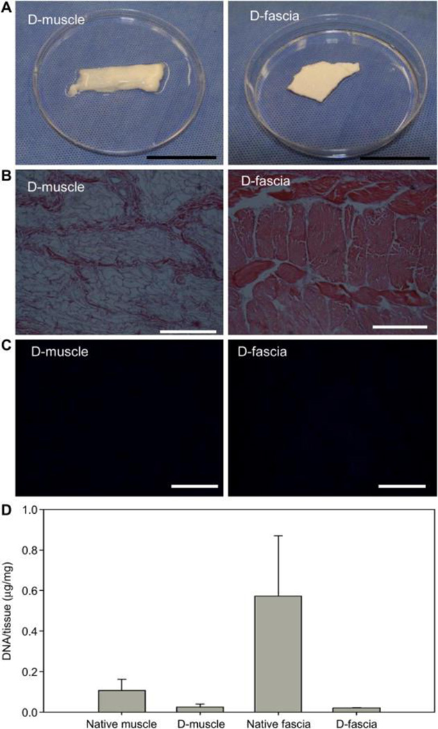
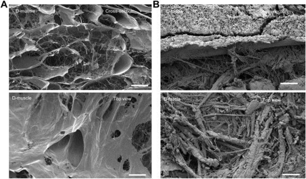
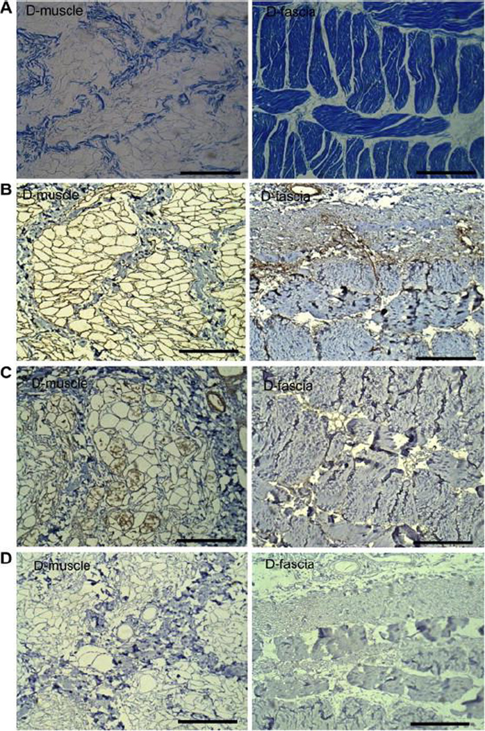
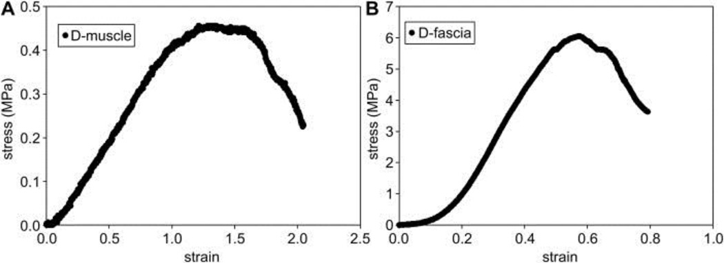
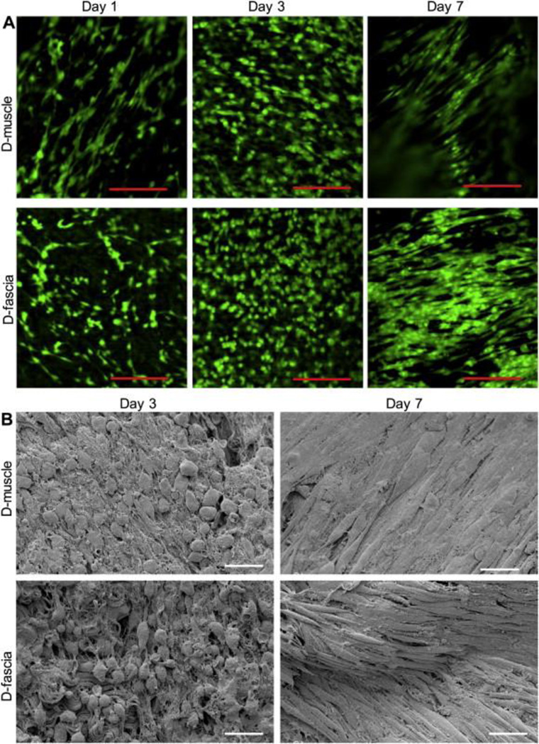
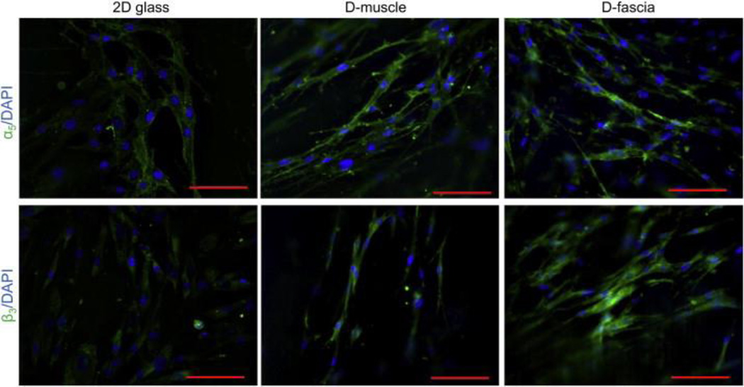
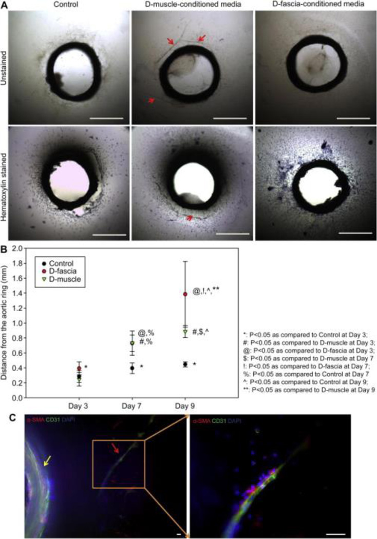
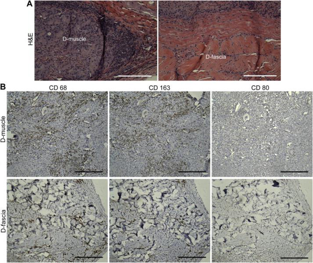
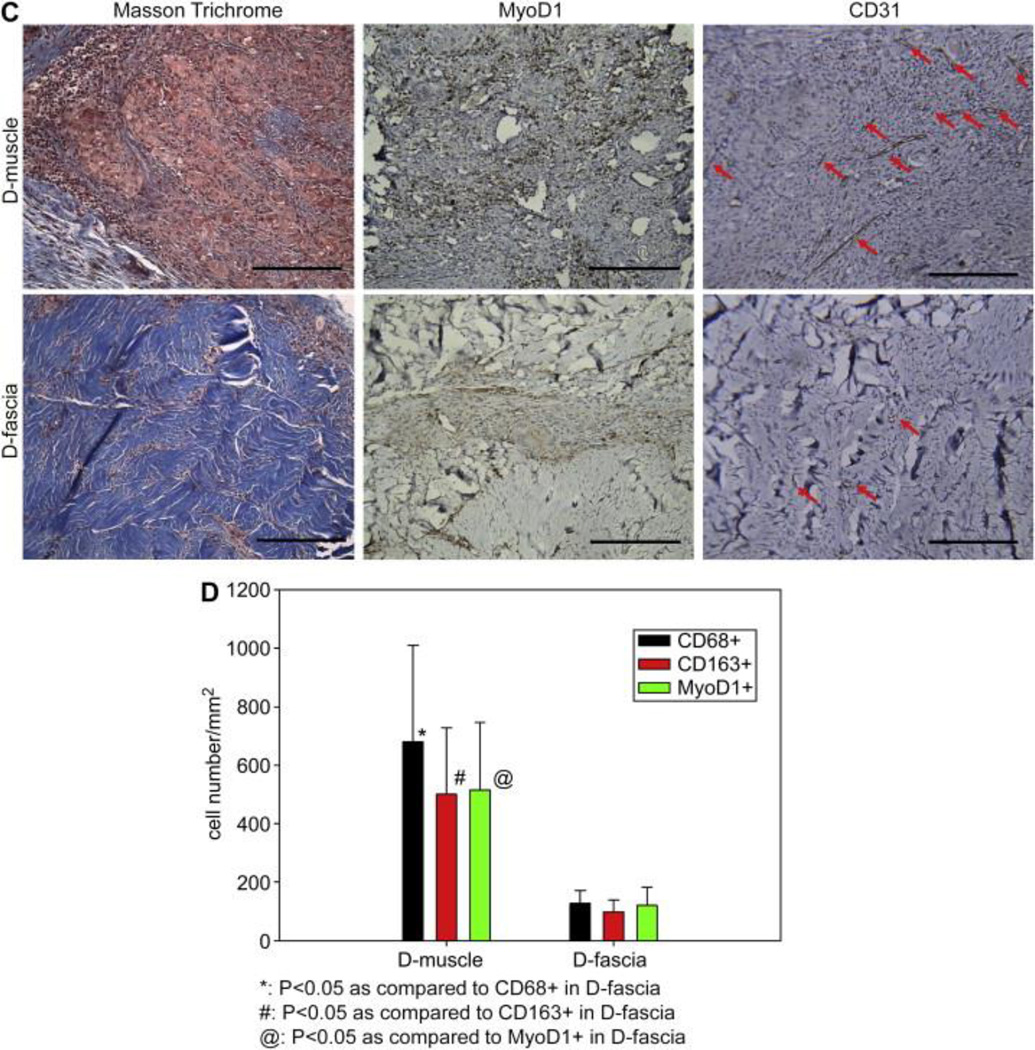
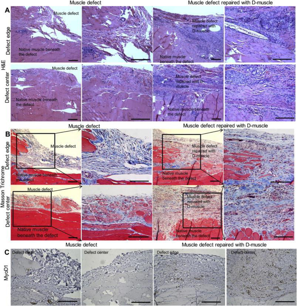
Ideal scaffolds that represent native extracellular matrix (ECM) properties of musculofascial tissues have great importance in musculofascial tissue engineering. However, detailed characterization of musculofascial tissues' ECM (particularly, of fascia) from large animals is still lacking. In this study, we developed a decellularization protocol for processing pig composite musculofascial tissues. Decellularized muscle (D-muscle) and decellularized fascia (D-fascia), which are two important components of decellularized musculofascial extracellular matrix (DMM), were comprehensively characterized. D-muscle and D-fascia retained intact three-dimensional architecture, strong mechanical properties, and bioactivity of compositions such as collagen, laminin, glycosaminoglycan, and vascular endothelial growth factor. D-muscle and D-fascia provided a compatible niche for human adipose-derived stem cell integration and proliferation. Heterotopic and orthotopic implantation of D-muscle and D-fascia in a rodent model further proved their biocompatibility and myogenic properties during the remodeling process. The differing characteristics of D-muscle from D-fascia (e.g. D-muscle's strong pro-angiogenic and pro-myogenic properties vs. D-fascia's strong mechanical properties) indicate different clinical application opportunities of D-muscle vs. D-fascia scaffolds. DMM comprising muscle and fascia ECM as a whole unit can thus provide not only a clinically translatable platform for musculofascial tissue repair and regeneration but also a useful standard for scaffold design in musculofascial tissue engineering.


Figures

Figure 1
Figure 1
Evaluation of cellular components’ removal from musculofascial tissues. (A) Images of muscle and fascial tissue after decellularization. (B) H&E staining of decellularized samples (D-muscle and D-fascia). (C) DAPI staining decellularized samples. (D) DNA content in dry weight samples. Scale bar = 20 mm in (A), 200 μm in (B) and (C).
Figure 2
Figure 2
SEM images of D-muscle (A) and D-fascia (B). The top row in each image shows the cross-sectional view, the bottom row shows the top view. Scale bar = 50 μm.
Figure 3
Figure 3
Characterization of D-muscle and D-fascia components. (A) Masson Trichrome staining shows the major structures of D-muscle and D-fascia are composed of collagen. (B) Laminin staining shows that after decellularization the fibrous structures among collagen structures are stained positively for laminin. Similar laminin distribution was noticed in D-fascia. (C) VEGF staining shows VEGF is retained in the nanofibrous structures within these small tubular structures in D-muscle. In D-fascia, VEGF staining was weakly positive in limited areas among collagen bundles (e.g. around blood vessels). (D) MHC-1 staining shows negative results in D-muscle and D-fascia. Scale bar = 200 μm.
Figure 4
Figure 4
Typical strain-stress curves of D-muscle (A) and D-fascia (B) measured by uniaxial tensile testing.
Figure 5
Figure 5
(A) Confocal images showing ASCs on D-muscle and D-fascia samples. ASCs were stained with Calcein AM on days 1, 3, and 7 after plating. (B) SEM images showing ASCs on D-muscle and D-fascia on days 3 and day 7 after integration. Scale bar = 100 μm in (A) and 50 μm in (B).
Figure 6
Figure 6
Confocal images showing ASCs on 2D glass slides, D-muscle, and D-fascia samples. ASCs were stained for integrin α5 or β3 (green) and DAPI (blue). Scale bar = 100 μm.
Figure 7
Figure 7
Aortic ring assay with D-muscle-conditioned media and D-fascia-conditioned media. (A) Phase contrast images of aortic rings on day 7 of in vitro cultures treated with control media (EGM media), D-muscle-conditioned media, and D-fascia-conditioned media. Samples were also stained with hematoxylin and imaged. Vessel branch outgrowth (as indicated by the red arrow) from the aortic ring in 3D collagen gel was observed on day 7 of in vitro culture treated with D-muscle-conditioned media. (B) Cell migration distance from the aortic ring measured on days 3, 7, and 9 of in vitro cultures treated with control media (EGM media), D-muscle-conditioned media, and D-fascia-conditioned media. (C) Confocal images showing vessel branch outgrowth (as indicated by the red arrow) from the aortic ring (as indicated by the yellow arrow) in 3D collagen gel on day 7 of in vitro culture treated with D-muscle-conditioned media. Samples were stained for α-smooth muscle actin (α-SMA, red), CD31 (green), and DAPI (blue). Scale bar = 1 mm in (A) and 50 μm in (C).
Figure 8
Figure 8
(A–C) Immunohistochemical staining of D-muscle and D-fascia 30 days after subcutaneous implantation in Fischer 344 rats. (A) H&E staining. (B) Staining for CD68, CD163, CD 80. (C) Masson Trichrome, MyoD1 and CD31 staining. Scale bar = 200 μm. (D) Summary of cell density (cells/mm2) of cells that stained positively for CD68, CD163 and MyoD1.
Figure 8
Figure 8
(A–C) Immunohistochemical staining of D-muscle and D-fascia 30 days after subcutaneous implantation in Fischer 344 rats. (A) H&E staining. (B) Staining for CD68, CD163, CD 80. (C) Masson Trichrome, MyoD1 and CD31 staining. Scale bar = 200 μm. (D) Summary of cell density (cells/mm2) of cells that stained positively for CD68, CD163 and MyoD1.
Figure 9
Figure 9
Immunohistochemical staining of muscle defect without repair and defect repaired with D-muscle on day 30 after surgery. (A) H&E staining. (B) Massion Trichrome staining. (C) MyoD1 staining. Scale bar = 200 μm.

Similar articles

Cited by

References

    1. Badylak SF, Weiss DJ, Caplan A, Macchiarini P. Engineered whole organs and complex tissues. Lancet. 2012;379(9819):943–952. - PubMed
    1. Deans TL, Elisseeff JH. Stem cells in musculoskeletal engineered tissue. Curr Opin Biotechnol. 2009;20(5):537–544. - PubMed
    1. Ma J, Holden K, Zhu J, Pan H, Li Y. The application of three-dimensional collagen-scaffolds seeded with myoblasts to repair skeletal muscle defects. J Biomed Biotechnol. 2011;2011:812135. - PMC - PubMed
    1. Merritt EK, Hammers DW, Tierney M, Suggs LJ, Walters TJ, Farrar RP. Functional assessment of skeletal muscle regeneration utilizing homologous extracellular matrix as scaffolding. Tissue Eng Part A. 2010;16(4):1395–1405. - PubMed
    1. Rossi CA, Pozzobon M, De Coppi P. Advances in musculoskeletal tissue engineering: moving towards therapy. Organogenesis. 2010;6(3):167–172. - PMC - PubMed

Publication types

MeSH terms