

Lead roles for supporting actors: critical functions of inner ear supporting cells
- PMID: 23347917
- PMCID: PMC3648608
- DOI: 10.1016/j.heares.2013.01.008
Lead roles for supporting actors: critical functions of inner ear supporting cells
Abstract
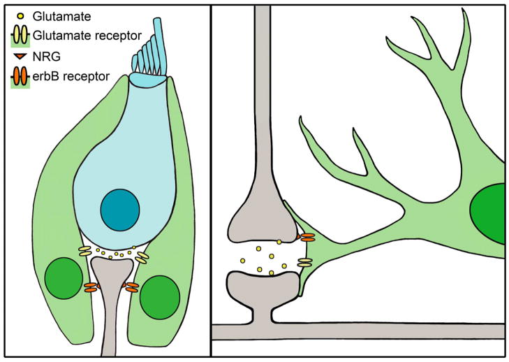
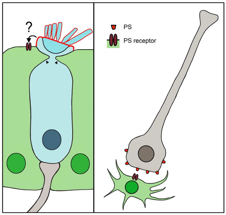
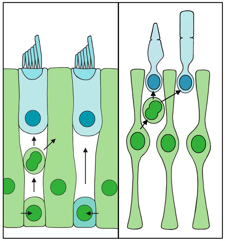
Many studies that aim to investigate the underlying mechanisms of hearing loss or balance disorders focus on the hair cells and spiral ganglion neurons of the inner ear. Fewer studies have examined the supporting cells that contact both of these cell types in the cochlea and vestibular end organs. While the roles of supporting cells are still being elucidated, emerging evidence indicates that they serve many functions vital to maintaining healthy populations of hair cells and spiral ganglion neurons. Here we review recent studies that highlight the critical roles supporting cells play in the development, function, survival, death, phagocytosis, and regeneration of other cell types within the inner ear. Many of these roles have also been described for glial cells in other parts of the nervous system, and lessons from these other systems continue to inform our understanding of supporting cell functions. This article is part of a Special Issue entitled "Annual Reviews 2013".
Keywords: BDNF; ERK 1/2; FGF; GLAST; HSP70; IHC; ISC; NRG; NT3; PS; SGN; T-cell restricted intracellular antigen-related protein; TEM; TIAR; brain-derived neurotrophic factor; extracellularly regulated kinases 1 and 2; fibroblast growth factor; glutamate aspartate transporter; heat shock protein 70; inner hair cell; inner supporting cell; neuregulin; neurotrophin-3; phosphatidylserine; spiral ganglion neuron; transmission electron microscopy.
Published by Elsevier B.V.
Figures
Similar articles
-
Fractalkine Signaling Regulates Macrophage Recruitment into the Cochlea and Promotes the Survival of Spiral Ganglion Neurons after Selective Hair Cell Lesion.J Neurosci. 2015 Nov 11;35(45):15050-61. doi: 10.1523/JNEUROSCI.2325-15.2015. J Neurosci. 2015. PMID: 26558776 Free PMC article.
-
Survival of adult spiral ganglion neurons requires erbB receptor signaling in the inner ear.J Neurosci. 2004 Oct 6;24(40):8651-61. doi: 10.1523/JNEUROSCI.0733-04.2004. J Neurosci. 2004. PMID: 15470130 Free PMC article.
-
Isolation of sphere-forming stem cells from the mouse inner ear.Methods Mol Biol. 2009;493:141-62. doi: 10.1007/978-1-59745-523-7_9. Methods Mol Biol. 2009. PMID: 18839346 Free PMC article.
-
The potential role of endogenous stem cells in regeneration of the inner ear.Hear Res. 2007 May;227(1-2):48-52. doi: 10.1016/j.heares.2006.12.015. Epub 2007 Jan 20. Hear Res. 2007. PMID: 17321086 Free PMC article. Review.
-
Hear, Hear for Notch: Control of Cell Fates in the Inner Ear by Notch Signaling.Biomolecules. 2020 Feb 28;10(3):370. doi: 10.3390/biom10030370. Biomolecules. 2020. PMID: 32121147 Free PMC article. Review.
Cited by
-
Conditional Ablation of Glucocorticoid and Mineralocorticoid Receptors from Cochlear Supporting Cells Reveals Their Differential Roles for Hearing Sensitivity and Dynamics of Recovery from Noise-Induced Hearing Loss.Int J Mol Sci. 2023 Feb 7;24(4):3320. doi: 10.3390/ijms24043320. Int J Mol Sci. 2023. PMID: 36834731 Free PMC article.
-
Genetic rescue of Muenke syndrome model hearing loss reveals prolonged FGF-dependent plasticity in cochlear supporting cell fates.Genes Dev. 2013 Nov 1;27(21):2320-31. doi: 10.1101/gad.228957.113. Epub 2013 Oct 21. Genes Dev. 2013. PMID: 24145799 Free PMC article.
-
Canonical Notch signaling plays an instructive role in auditory supporting cell development.Sci Rep. 2016 Jan 20;6:19484. doi: 10.1038/srep19484. Sci Rep. 2016. PMID: 26786414 Free PMC article.
-
Delivery of Adeno-Associated Virus Vectors in Adult Mammalian Inner-Ear Cell Subtypes Without Auditory Dysfunction.Hum Gene Ther. 2018 Apr;29(4):492-506. doi: 10.1089/hum.2017.120. Epub 2018 Jan 22. Hum Gene Ther. 2018. PMID: 29130354 Free PMC article.
-
Gap junctional coupling is essential for epithelial repair in the avian cochlea.J Neurosci. 2014 Nov 26;34(48):15851-60. doi: 10.1523/JNEUROSCI.1932-14.2014. J Neurosci. 2014. PMID: 25429127 Free PMC article.
References
-
- Aboitiz F, Montiel J, Morales D, Concha M. Evolutionary divergence of the reptilian and the mammalian brains: considerations on connectivity and development. Brain Res Brain Res Rev. 2002;39:141–153. - PubMed
-
- Ahmad M, Bohne BA, Harding GW. An in vivo tracer study of noise-induced damage to the reticular lamina. Hear Res. 2003;175:82–100. - PubMed
-
- Baird RA, Steyger PS, Schuff NR. Mitotic and nonmitotic hair cell regeneration in the bullfrog vestibular otolith organs. Ann N Y Acad Sci. 1996;781:59–70. - PubMed
Publication types
MeSH terms
Grants and funding
LinkOut - more resources
Full Text Sources
Other Literature Sources
Miscellaneous
