. 2013 May;30(5):1015-31.
doi: 10.1093/molbev/mst014. Epub 2013 Jan 24.

Genesis and regulatory wiring of retroelement-derived domesticated genes: a phylogenomic perspective

Affiliations

Genesis and regulatory wiring of retroelement-derived domesticated genes: a phylogenomic perspective

Janez Kokošar et al. Mol Biol Evol. 2013 May.

Abstract

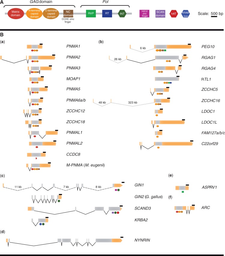
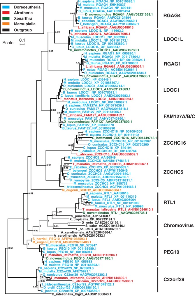
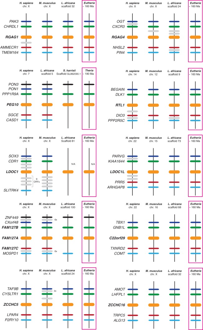
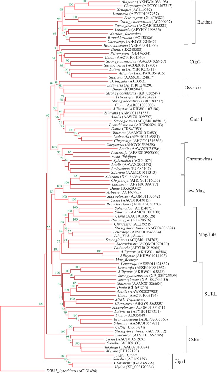
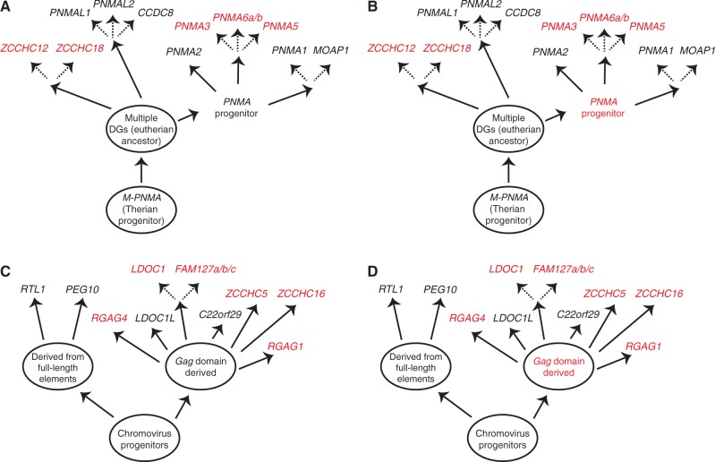
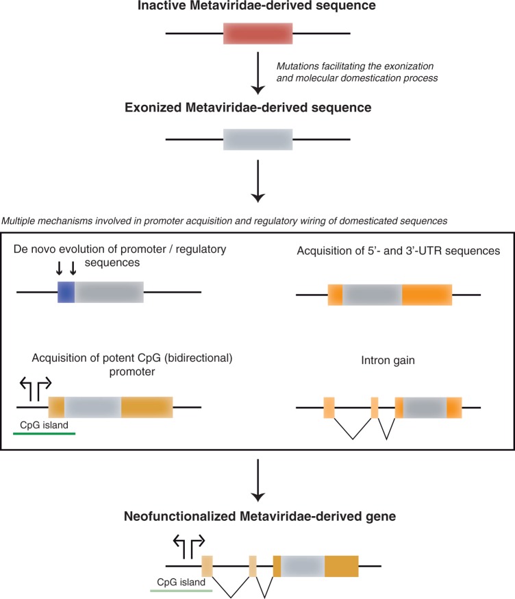
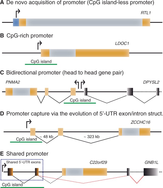
Molecular domestications of transposable elements have occurred repeatedly during the evolution of eukaryotes. Vertebrates, especially mammals, possess numerous single copy domesticated genes (DGs) that have originated from the intronless multicopy transposable elements. However, the origin and evolution of the retroelement-derived DGs (RDDGs) that originated from Metaviridae has been only partially elucidated, due to absence of genome data or to limited analysis of a single family of DGs. We traced the genesis and regulatory wiring of the Metaviridae-derived DGs through phylogenomic analysis, using whole-genome information from more than 90 chordate genomes. Phylogenomic analysis of these DGs in chordate genomes provided direct evidence that major diversification has occurred in the ancestor of placental mammals. Mammalian RDDGs have been shown to originate in several steps by independent domestication events and to diversify later by gene duplications. Analysis of syntenic loci has shown that diverse RDDGs and their chromosomal positions were fully established in the ancestor of placental mammals. By analysis of active Metaviridae lineages in amniotes, we have demonstrated that RDDGs originated from retroelement remains. The chromosomal gene movements of RDDGs were highly dynamic only in the ancestor of placental mammals. During the domestication process, de novo acquisition of regulatory regions is shown to be a prerequisite for the survival of the DGs. The origin and evolution of de novo acquired promoters and untranslated regions in diverse mammalian RDDGs have been explained by comparative analysis of orthologous gene loci. The origin of placental mammal-specific innovations and adaptations, such as placenta and newly evolved brain functions, was most probably connected to the regulatory wiring of DGs and their rapid fixation in the ancestor of placental mammals.


Figures

F<sc>ig</sc>. 1.
Fig. 1.
Gene structures of human RDDGs. (A) Color-coded protein domains of Metaviridae retroelements and additional domains that are present in human RDDG genes. (B) Exon–intron organization of human RDDG genes. Exons are shown as boxes and introns as connecting lines. Gray regions of exons denote the protein-coding sequences, whereas UTRs are represented as orange boxes. Protein domains that are present in RDDG genes are shown schematically below the protein-coding sequences. (a) PNMA family; (b) sushi/Chromovirus family; (c) integrase-derived RDDG genes; (d) NYNRIN gene; (e) retroelement protease-derived ASPRV1 gene; and (f) ARC gene. Scale bar represents 500 bp. Distances between exons are drawn to scale except in the cases of extremely long introns. RVP, protease; RT, reverse transcriptase; INT, integrase.
F<sc>ig</sc>. 2.
Fig. 2.
Bayesian phylogeny of the sushi family of RDDGs. The tree was inferred by MrBayes 3 program under a Poisson + G4 model from the N-terminal capsid of gag domain. Only posterior probabilities larger than 0.5 are shown. The scale bar corresponds to 0.1 substitutions per site. The N-terminal capsid of gag domain from Ciona intestinalis Cigr2 retroelement was used to root this tree.
F<sc>ig</sc>. 3.
Fig. 3.
Conserved synteny in the sushi family of RDDGs. Chromosomal regions carrying all the RDDGs in the species considered in this analysis were compared, and neighbouring genes with conserved synteny were identified. Horizontal lines denote orthologous relationships. Each RDDG gene is represented in bold as a horizontal orange line on the chromosome. Neighbouring genes that are in synteny are shown with a schematic indication of their distance (not to scale). Ancestral states of the RDDG chromosomal positions were reconstructed from comparison of syntenic positions between multiple mammalian lineages.
F<sc>ig</sc>. 4.
Fig. 4.
Tracing the RDDGs progenitors in Amniota. The rooted NJ tree was inferred by MEGA 5.0 program under a p-distance + pairwise deletion model from the combined Metaviridae RT and RNAse H domains. Reliability for the internal branches was assessed using the 1,000 bootstrap replications; nodes with confidence values greater than 50% are indicated. The DIRS1 element from Lytechinus (AC131494) has been used to root this tree. Most sequences were obtained from the GenBank; genus names and accession numbers are included.
F<sc>ig</sc>. 5.
Fig. 5.
Chromosomal gene movements of RDDGs occurred only in the ancestor of placental mammals. The putative ancestral chromosomal locations and potential cases of movement of RDDGs from autosomes to the X-chromosome, or from the X-chromosome to autosomes, were inferred from our phylogenetic and conserved synteny analyses. (A, B) Schematic representations of alternative hypotheses regarding the movement of PNMA family genes from the autosomes to the X-chromosome or from the X-chromosome to the autosomes. The therian PNMA gene progenitor is located on the autosome. A few independent originations from this progenitor may generate autosomal and X-chromosome (genes located on the X-chromosome are marked in red) gene movements. In the case of PNMA progenitor, the ancestral chromosomal position is unknown and can be either autosomal (scenario under hypothesis A) or on the X chromosome (scenario under hypothesis B). (C, D) Schematic representation of alternative hypotheses regarding the movement of the sushi family genes from the autosomes to the X-chromosome or from the X-chromosome to autosomes. The chromoviral full-length progenitor was most probably located on the autosome, because the RTL1 and PEG10 genes are also autosomal. Numerous independent originations from the chromoviral gag progenitor may generate autosomal and X-chromosome (genes located on the X-chromosome are marked in red) movements. In the case of the chromoviral gag progenitor, the ancestral chromosomal position is unknown and can be either autosomal (scenario under hypothesis C) or on the X chromosome (scenario under hypothesis D).
F<sc>ig</sc>. 6.
Fig. 6.
Mechanisms involved in the process of RDDG neofunctionalization. In the transition phase from retroelement remains to the first RDDGs, many nucleotide changes were necessary for the neofunctionalization. One of the crucial steps in the process of neofunctionalization was the exonization of retroelement domains (gag, protease, and integrase), which produced ready-to-use modules. Retroelement remains in mammalian genomes will normally turn into pseudogenes, due to lack of a promoter, and they can survive as a functional gene only if they recruit a new promoter sequence. To become expressed at a significant level and in the tissues where it can exert a selectively beneficial function, a new gene needs to acquire a core promoter and other structural elements that regulate its expression. Exons and introns are shown as orange (5'- and 3'-UTR regions) or gray (coding part of the exons) boxes and connecting lines. A de novo acquired promoter is shown in blue.
F<sc>ig</sc>. 7.
Fig. 7.
Diverse sources of RDDGs promoters. Various scenarios that lead to the transcription of RDDG gene copies are illustrated. (A) Recruitment of proto-promoters from the CpG island-less region. (B) Recruitment of proto-promoters from the CpG-rich island. (C) Recruitment of a bidirectional (CpG-enriched) promoter from neighboring gene in the vicinity of the RDDG gene. (D) Recruitment of distant promoters in the genomic neighbourhood via the acquisition of a new 5'-untranslated exon–intron structure. (E) Sharing of the unidirectional (CpG-enriched) promoter from a neighboring gene in the vicinity of the RDDG gene. Exons and introns are represented by orange and gray (RDDG genes) or black (neighbouring genes in the case of bidirectional promoters) boxes and connecting lines. Distances between exons are not to scale.

References

    1. Almeida LM, Silva IT, Silva WA, Jr, Castro JP, Riggs PK, Carareto CM, Amaral ME. The contribution of transposable elements to Bos taurus gene structure. Gene. 2007;390:180–189. - PubMed
    1. Andreassi C, Riccio A. To localize or not to localize: mRNA fate is in 3'UTR ends. Trends Cell Biol. 2009;19:465–474. - PubMed
    1. Bao W, Kapitonov VV, Jurka J. Ginger DNA transposons in eukaryotes and their evolutionary relationships with long terminal repeat retrotransposons. Mob DNA. 2010;1:3. - PMC - PubMed
    1. Barrett LW, Fletcher S, Wilton SD. Regulation of eukaryotic gene expression by the untranslated gene regions and other non-coding elements. Cell Mol Life Sci. 2012;69: 3613–3634. - PMC - PubMed
    1. Benton MJ, Twitchett RJ. How to kill (almost) all life: the end-Permian extinction event. Trends Ecol Evol. 2003;18:358–365.

Publication types

LinkOut - more resources