

The male mammary gland: a target for the xenoestrogen bisphenol A
- PMID: 23348055
- PMCID: PMC3998714
- DOI: 10.1016/j.reprotox.2013.01.002
The male mammary gland: a target for the xenoestrogen bisphenol A
Abstract
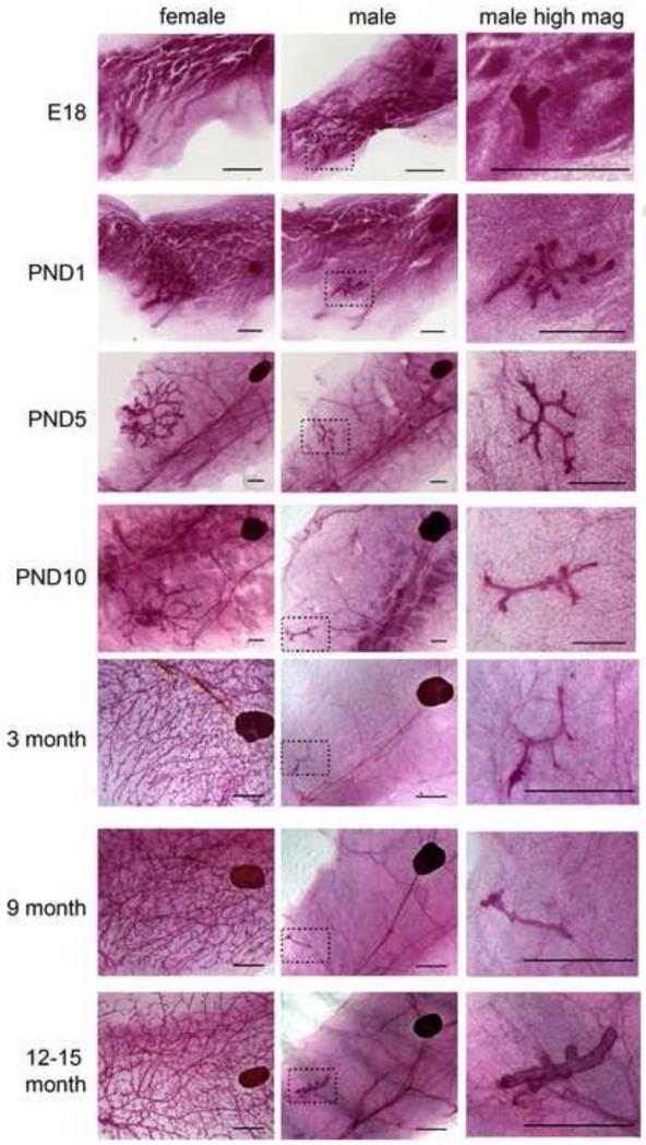
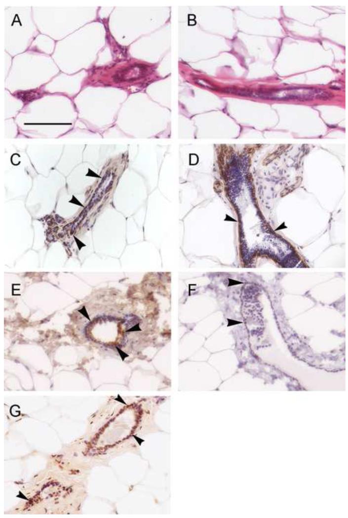
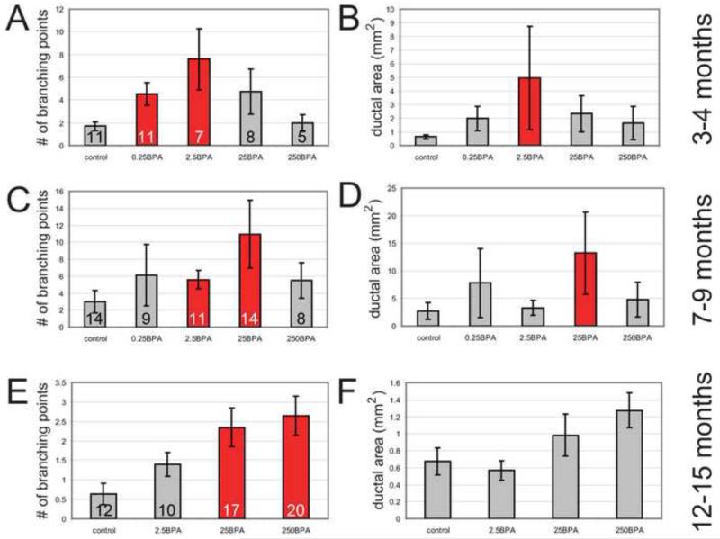
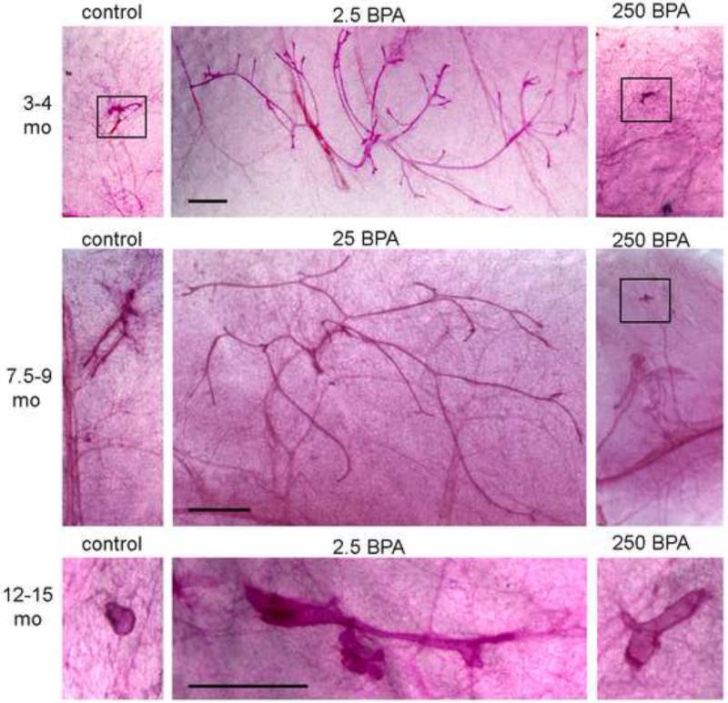
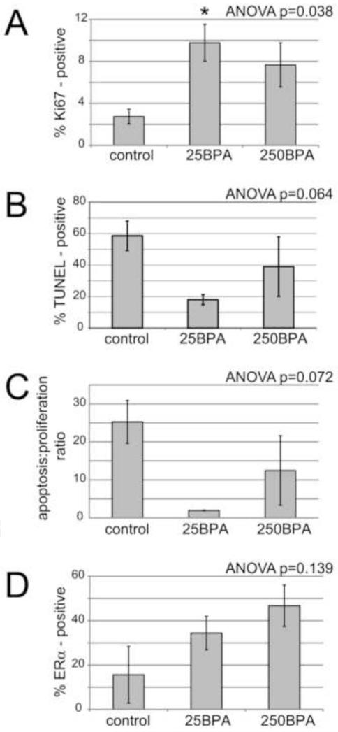
Males of some strains of mice retain their mammary epithelium even in the absence of nipples. Here, we have characterized the mammary gland in male CD-1 mice both in whole mounts and histological sections. We also examined the effects of bisphenol A (BPA), an estrogen mimic that alters development of the female mouse mammary gland. BPA was administered at a range of environmentally relevant doses (0.25-250μg/kg/day) to pregnant and lactating mice and then the mammary glands of male offspring were examined at several periods in adulthood. We observed age- and dose-specific effects on mammary gland morphology, indicating that perinatal BPA exposures alter the male mammary gland in adulthood. These results may provide insight into gynecomastia, the most common male breast disease in humans, where proliferation of the mammary epithelium leads to breast enlargement.
Copyright © 2013 Elsevier Inc. All rights reserved.
Figures
Similar articles
-
Bisphenol S alters development of the male mouse mammary gland and sensitizes it to a peripubertal estrogen challenge.Toxicology. 2019 Aug 1;424:152234. doi: 10.1016/j.tox.2019.06.005. Epub 2019 Jun 12. Toxicology. 2019. PMID: 31201878 Free PMC article.
-
Estrogens in the wrong place at the wrong time: Fetal BPA exposure and mammary cancer.Reprod Toxicol. 2015 Jul;54:58-65. doi: 10.1016/j.reprotox.2014.09.012. Epub 2014 Sep 30. Reprod Toxicol. 2015. PMID: 25277313 Free PMC article. Review.
-
Low-dose BPA exposure alters the mesenchymal and epithelial transcriptomes of the mouse fetal mammary gland.PLoS One. 2013 May 21;8(5):e63902. doi: 10.1371/journal.pone.0063902. Print 2013. PLoS One. 2013. PMID: 23704952 Free PMC article.
-
Bisphenol A alters the development of the rhesus monkey mammary gland.Proc Natl Acad Sci U S A. 2012 May 22;109(21):8190-5. doi: 10.1073/pnas.1120488109. Epub 2012 May 7. Proc Natl Acad Sci U S A. 2012. PMID: 22566636 Free PMC article.
-
Alteration of mammary gland development by bisphenol a and evidence of a mode of action mediated through endocrine disruption.Mol Cell Endocrinol. 2018 Nov 5;475:29-53. doi: 10.1016/j.mce.2018.06.015. Epub 2018 Jul 23. Mol Cell Endocrinol. 2018. PMID: 30048677 Review.
Cited by
-
Potential Mechanisms of Bisphenol A (BPA) Contributing to Human Disease.Int J Mol Sci. 2020 Aug 11;21(16):5761. doi: 10.3390/ijms21165761. Int J Mol Sci. 2020. PMID: 32796699 Free PMC article. Review.
-
Bisphenol S alters development of the male mouse mammary gland and sensitizes it to a peripubertal estrogen challenge.Toxicology. 2019 Aug 1;424:152234. doi: 10.1016/j.tox.2019.06.005. Epub 2019 Jun 12. Toxicology. 2019. PMID: 31201878 Free PMC article.
-
Estrogens in the wrong place at the wrong time: Fetal BPA exposure and mammary cancer.Reprod Toxicol. 2015 Jul;54:58-65. doi: 10.1016/j.reprotox.2014.09.012. Epub 2014 Sep 30. Reprod Toxicol. 2015. PMID: 25277313 Free PMC article. Review.
-
Adipose stem cells are sexually dimorphic cells with dual roles as preadipocytes and resident fibroblasts.Nat Commun. 2024 Sep 2;15(1):7643. doi: 10.1038/s41467-024-51867-9. Nat Commun. 2024. PMID: 39223126 Free PMC article.
-
Visceral obesity and cell cycle pathways serve as links in the association between bisphenol A exposure and breast cancer.Oncol Lett. 2020 Jul;20(1):33-42. doi: 10.3892/ol.2020.11553. Epub 2020 Apr 21. Oncol Lett. 2020. PMID: 32565931 Free PMC article.
References
-
- Hennighausen L, Robinson GW. Think globally, act locally: the making of a mouse mammary gland. Genes Dev. 1998;12:449–55. - PubMed
-
- Iuanow E, Kettler M, Slanetz PJ. Spectrum of disease in the male breast. AJR Am J Roentgenol. 2011;196:W247–59. - PubMed
-
- Markey CM, Coombs MA, Sonnenschein C, Soto AM. Mammalian development in a changing environment: exposure to endocrine disruptors reveals the developmental plasticity of steroid-hormone target organs. Evolution and Development. 2003;5:67–75. - PubMed
Publication types
MeSH terms
Substances
Grants and funding
LinkOut - more resources
Full Text Sources
Other Literature Sources
