

Renal medullary carcinoma: molecular, immunohistochemistry, and morphologic correlation
- PMID: 23348212
- PMCID: PMC7604824
- DOI: 10.1097/PAS.0b013e3182770406
Renal medullary carcinoma: molecular, immunohistochemistry, and morphologic correlation
Abstract
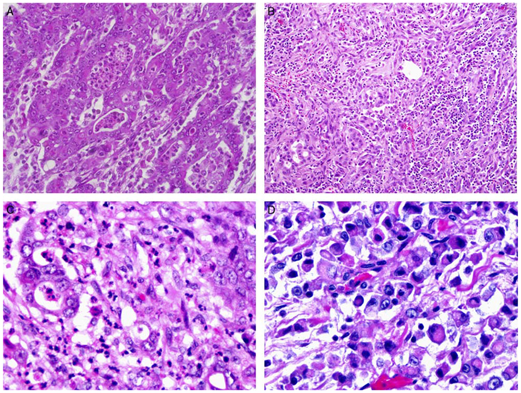
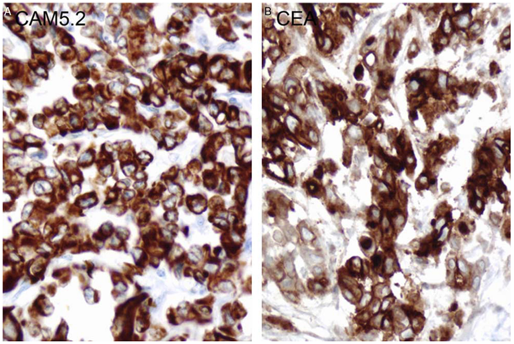
Renal medullary carcinoma, a highly aggressive tumor mainly occurring in patients with sickle cell hemoglobinopathy, is characterized by advanced stage at the time of presentation and poor response to treatment. Currently, the pathogenesis of this tumor is not well understood. In this study, the clinicopathologic features and molecular changes of 15 renal medullary carcinoma cases were evaluated. These cases demonstrated male predominance (M:F=2:1) with a median age of 26 years. The tumors occurred predominantly in the right kidney with an average size of 5.9 cm. Immunohistochemistry analysis showed that the neoplastic cells were positive for CEA (7/8), AE1/3 (8/8), CAM5.2 (7/7), CK7 (5/5), CK20 (4/6), and vimentin (6/6). Absence of SMARCB1 protein expression in tumor cells was demonstrated in all of the 7 cases analyzed. By polymerase chain reaction-based microsatellite analysis, loss of heterozygosity of SMARCB1 was identified in 9 of 10 cases. These data suggest that inactivation of SMARCB1 may play a role in the pathogenesis of renal medullary carcinoma.
Figures


References
-
- Davis CJ Jr, Mostofi FK, Sesterhenn IA. Renal medullary carcinoma. The seventh sickle cell nephropathy. Am J Surg Pathol. 1995;19:1–11. - PubMed
-
- Medjkane S, Novikov E, Versteege I. et al. The tumor suppressor hSNF5/INI1 modulates cell growth and actin cytoskeleton organization. Cancer Res. 2004;64:3406–3413. - PubMed
-
- Versteege I, Medjkane S, Rouillard D. et al. A key role of the hSNF5/INI1 tumour suppressor in the control of the G1-S transition of the cell cycle. Oncogene. 2002;21:6403–6412. - PubMed
Publication types
MeSH terms
Substances
Grants and funding
LinkOut - more resources
Full Text Sources
Other Literature Sources
Medical
Research Materials
Miscellaneous
