Comparative Study
. 2013 Feb;8(2):373-92.
doi: 10.1038/nprot.2013.001. Epub 2013 Jan 24.

Incorporation of a viral DNA-packaging motor channel in lipid bilayers for real-time, single-molecule sensing of chemicals and double-stranded DNA

Affiliations
Comparative Study

Incorporation of a viral DNA-packaging motor channel in lipid bilayers for real-time, single-molecule sensing of chemicals and double-stranded DNA

Farzin Haque et al. Nat Protoc. 2013 Feb.

Abstract

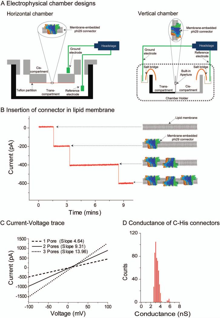
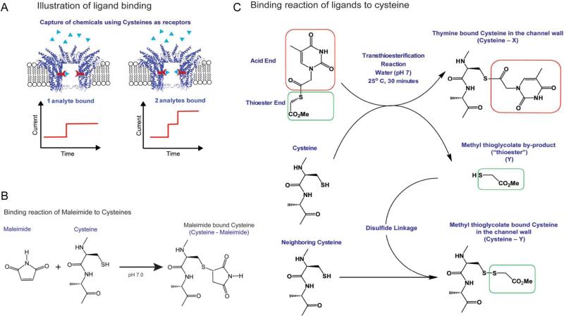
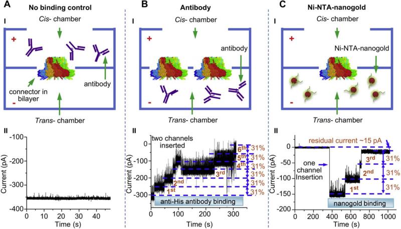
Over the past decade, nanopores have rapidly emerged as stochastic biosensors. This protocol describes the cloning, expression and purification of the channel of the bacteriophage phi29 DNA-packaging nanomotor and its subsequent incorporation into lipid membranes for single-pore sensing of double-stranded DNA (dsDNA) and chemicals. The membrane-embedded phi29 nanochannel remains functional and structurally intact under a range of conditions. When ions and macromolecules translocate through this nanochannel, reliable fingerprint changes in conductance are observed. Compared with other well-studied biological pores, the phi29 nanochannel has a larger cross-sectional area, which enables the translocation of dsDNA. Furthermore, specific amino acids can be introduced by site-directed mutagenesis within the large cavity of the channel to conjugate receptors that are able to bind specific ligands or analytes for desired applications. The lipid membrane-embedded nanochannel system has immense potential nanotechnological and biomedical applications in bioreactors, environmental sensing, drug monitoring, controlled drug delivery, early disease diagnosis and high-throughput DNA sequencing. The total time required for completing one round of this protocol is around 1 month.


Figures

Figure 1
Figure 1. Structure of phi29 DNA packaging motor and connector nanochannel
(a, b) An illustration of DNA translocation through the phi29 DNA packaging motor. (c) Side view of the phi29 connector showing the acidic (red), basic (blue), and other (white) amino acids. (d) Top view of the connector showing the diameter of the narrow part and wide part of the channel. Figures reproduced with permissions from: (a) Ref. , © American Chemical Society; (bd) Ref. , © Nature Publishing Group.
Figure 2
Figure 2. Example of plasmid construction for over-expression of connectors with C-terminal modification by two-step PCR.
(a) The linker was attached to the 3’-end of GP10 gene in the first PCR by a primer pair F1-R1. In a second PCR, amino acids were incorporated downstream using primer pair F1-R2, which contained NdeI and XhoI restriction sites, respectively. (b) The second PCR product was digested with both NdeI and XhoI, an ligated into the NdeI/XhoI sites of the vector pET-21a(+). (c) Sequences of primers. (d) Summary of the connector protein extension constructs. Underlined sequence represents TEV (Tobacco Etch Virus) protease recognition sequence; denotes the TEV cleavage site. Figures reproduced with permissions from: (ac) Ref. , © Nature Publishing Group.
Figure 3
Figure 3. Images of fluorescently labeled liposomes containing the connector
(a) Epifluorescence images of liposome: lipid labeled with NBD-PE without connector (left); a proteoliposomes reconstituted by FITC-labeled connectors (middle); FITC-connector mixed non-specifically with liposomes (right). (b) Separation of liposome/FITC-connector complexes by sucrose gradient sedimentation. Free connectors appeared in the top fractions while proteoliposomes remained in the lower fractions. Fractions 1–12 are not shown. Figures reproduced with permissions from Ref. , © Nature Publishing Group.
Figure 4
Figure 4. Insertion of connector channels in the planar lipid membrane
(a) Schematic representation of electrophysiological assay setup: horizontal (left) and vertical (right) chambers. Note: figure not drawn to scale. (b) Insertion of the re-engineered phi29 connector into lipid membrane and demonstration of robust conductivity property. Discrete steps of current representing the insertion of one connector for each step (left) and illustration (right). Connector insertion into the bilayer only occurred when connector-reconstituted proteoliposomes were fused into the bilayer. Note that the orientation of the connector channel is random. (c) Data demonstrating robust properties of connector indicated by strong linear I–V relationship. Increased number of connectors is associated with larger slopes but identical conductance per channel. (d) Histogram showing uniform conductance of connector channels in the membrane. Figures reproduced with permissions from: (cd) Ref. , © The Royal Society of Chemistry.
Figure 5
Figure 5. Translocation of dsDNA through membrane-embedded connector channel
(a) Illustration of dsDNA translocation in vitro; (b) Translocation of linear dsDNA induced numerous partial current blockages. Insert: Magnified image of a current blockage event caused by the translocation of a single DNA molecule. (c) Histogram of current blockage percentage induced by linear dsDNA (2 kbp) translocations with a total 3,264 events in the presence of 1M NaCl, pH 7.8 under (–)75 mV constant potential. (d) Comparison of dwell times for translocation of 38 bp and 5.5 kbp dsDNA. (eg) DNA translocation events through a single connector channel observed under a ramping potential without DNA (e) and with dsDNA in both cis and trans compartments (f) and (g). Data prove the one-directional trafficking of dsDNA across the channel. Figures reproduced with permissions from ref. (a,e-g); ref. (c); ref. (d).
Figure 6
Figure 6. Quantitative PCR (Q-PCR) analysis of DNA translocation events
(a) Q-PCR amplification curves of the dilution series run in triplicate (top) using SYBR Green dye, which binds to dsDNA. The threshold level (set in the exponential amplification phase) is the detection point at which the PCR reaction reaches a fluorescent intensity above background. The cycle at which the DNA sample reaches this threshold level is called the Threshold Cycle (CT); A standard curve with the CT plotted against the log of the starting quantity of template for each dilution (bottom). (b) Quantitative analysis of the total number of DNA passing through one of the connectors in the lipid membrane from the trans compartment to the cis compartment (top). Negative controls (bottom) were carried out under the same condition but without connectors. The error bars represent standard deviations of the mean from nine independent experiments and four negative control experiments. (c) Plot of the total number of DNA passing through multiple connectors in the lipid membrane (noted on the right). (d) Plot of the total number of DNA molecules translocated under membrane leaking conditions over time without any connector channels in the membrane. (e) A comparison of copy number of translocated dsDNA measured by Q-PCR with the relevant blockage events counted from current trace recorded in two independent experiments (Note: Only the events with 29–34% current blockages were counted). The error bars represent the standard deviation of three independent Q-PCR measurements. Figures reproduced with permissions from: (ad) Ref. , © Nature Publishing Group; (e) Ref. , © The Royal Society of Chemistry.
Figure 7
Figure 7. Conjugation of chemical ligands to channel wall
(a) Schematic of ligand binding to the inner wall of the connector pore which will result in the reduction of channel size as indicated by uniform stepwise blockage of channel current. (b) Reaction scheme of maleimidecysteine reaction. (c) Transthioesterification method, showing that cysteines are readily modified with nucleobases when exposed to thioesters, using thymine thioester as an example; a second reaction via disulfide linkage results in the binding of the methyl thioglycolate by-product. Figures reproduced with permissions from: Ref. , © American Chemical Society.
Figure 8
Figure 8. Capture and fingerprinting of chemicals in the channel lumen
(a) Data showing discrete blocking steps due to the binding of thioesters groups containing ethane to accessible cysteine residues introduced by mutagenesis in the channel wall. Insert: Magnified current trace showing the transient and permanent current blockage events. (b) Analysis of current blockage events induced by the binding of thioesters to cysteine residues located in the channel wall. Histogram of permanent binding events for the binding of thioesters groups containing ethane, thymine, and benzene respectively. Figures reproduced with permissions from: Ref. , © American Chemical Society.
Figure 9
Figure 9. Capture and fingerprinting of antibodies or chemical ligand at the C-terminal end of the channel
(a) Negative control. Since the antibody was placed in the cis compartment opposite to the C-terminal, no antibody binding occurs and no changes in current are observed. (b) Six discrete steps of changes for two C-His-tagged connector channels induced by anti-His tag antibodies. (c) Three discrete steps of changes for one C-His-tagged connector channels induced by Ni-NTA nanogold binding (1.8 nm). Figures reproduced with permissions from: Ref., © Elseiver.

Similar articles

Cited by

References

    1. Guo P, Grimes S, Anderson D. A defined system for in vitro packaging of DNA-gp3 of the Bacillus subtilis bacteriophage phi29. Proc. Natl. Acad. Sci. USA. 1986;83:3505–3509. - PMC - PubMed
    1. Smith DE, et al. The bacteriophage phi29 portal motor can package DNA against a large internal force. Nature. 2001;413:748–752. - PubMed
    1. Rickgauer JP, et al. Portal motor velocity and internal force resisting viral DNA packaging in bacteriophage phi 29. Biophysical Journal. 2008;94:159–167. - PMC - PubMed
    1. Guo P, Erickson S, Anderson D. A small viral RNA is required for in vitro packaging of bacteriophage phi29 DNA. Science. 1987;236:690–694. - PubMed
    1. Lee CS, Guo P. In vitro assembly of infectious virions of ds-DNA phage f29 from cloned gene products and synthetic nucleic acids. J. Virol. 1995;69:5018–5023. - PMC - PubMed

Publication types

MeSH terms