

Chromatin structure outside and inside the nucleus
- PMID: 23348669
- PMCID: PMC3557801
- DOI: 10.1002/bip.22157
Chromatin structure outside and inside the nucleus
Abstract
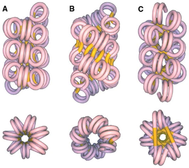
The structure of the 30-nm chromatin fiber has provided, over the years, an important reference in chromatin studies. Originally derived from electron microscopic studies of soluble chromatin fibers released by restriction digestion, the gross structural features of such fragments have been supported by biophysical methods such as low angle X-ray and neutron scattering, sedimentation, light scattering, and electric dichroism. Electron microscopy and sedimentation velocity measurements demonstrated that reconstituted chromatin fibers, prepared from repeating arrays of high affinity nucleosome positioning sequences, retain the same overall features as observed for native chromatin fibers. It had been suggested that the 30 nm fiber might be the form assumed in vivo by transcriptionally silent chromatin, but individual gene or genome-wide studies of chromatin released from nuclei do not reveal any such simple correlation. Furthermore, even though the 30 nm fiber has been thought to represent an intermediate in the hierarchical folding of DNA into chromosomes, most analyses of chromatin folding within the nucleus do not detect any regular extended compact structures. However, there are important exceptions in chicken erythroid cell nuclei as well as in transcribed regions that form extended loops. Localized domains within the nucleus, either at the surface of chromosome domains or constrained as a specialized kind of constitutive heterochromatin by specific DNA binding proteins, may adopt 30 nm fiber-like structures.
Copyright © 2012 Wiley Periodicals, Inc.
Figures
References
Publication types
MeSH terms
Substances
Grants and funding
LinkOut - more resources
Full Text Sources
Other Literature Sources
