. 2013 Feb;123(2):908-16.
doi: 10.1172/JCI65154. Epub 2013 Jan 25.

Platelet ITAM signaling is critical for vascular integrity in inflammation

Affiliations

Platelet ITAM signaling is critical for vascular integrity in inflammation

Yacine Boulaftali et al. J Clin Invest. 2013 Feb.

Abstract

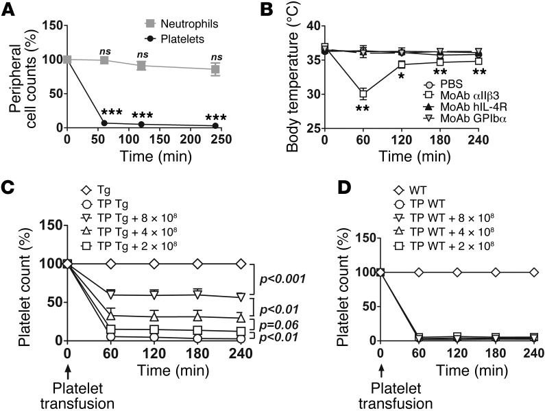
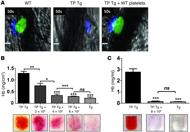
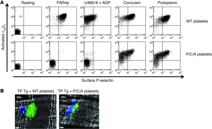
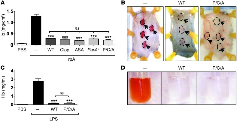
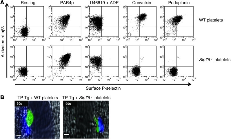
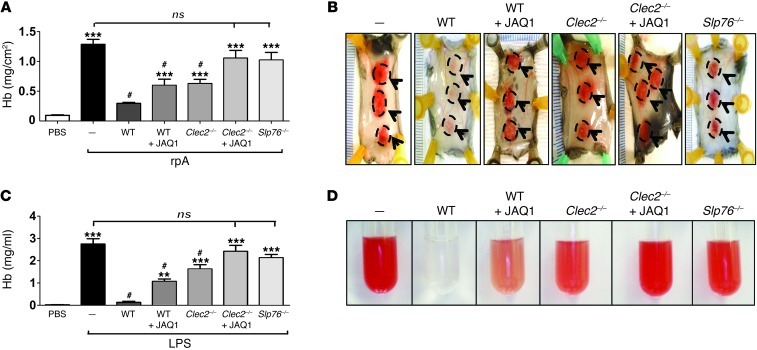
Platelets play a critical role in maintaining vascular integrity during inflammation, but little is known about the underlying molecular mechanisms. Here we report that platelet immunoreceptor tyrosine activation motif (ITAM) signaling, but not GPCR signaling, is critical for the prevention of inflammation-induced hemorrhage. To generate mice with partial or complete defects in these signaling pathways, we developed a protocol for adoptive transfer of genetically and/or chemically inhibited platelets into thrombocytopenic (TP) mice. Unexpectedly, platelets with impaired GPCR signaling, a crucial component of platelet plug formation and hemostasis, were indistinguishable from WT platelets in their ability to prevent hemorrhage at sites of inflammation. In contrast, inhibition of GPVI or genetic deletion of Clec2, the only ITAM receptors expressed on mouse platelets, significantly reduced the ability of platelets to prevent inflammation-induced hemorrhage. Moreover, transfusion of platelets without ITAM receptor function or platelets lacking the adapter protein SLP-76 into TP mice had no significant effect on vascular integrity during inflammation. These results indicate that the control of vascular integrity is a major function of immune-type receptors in platelets, highlighting a potential clinical complication of novel antithrombotic agents directed toward the ITAM signaling pathway.


Figures

Figure 1
Figure 1. Adoptive transfer of WT platelets into TP hIL-4Rα/GPIbα–Tg mice.
(A) TP was induced in hIL-4Rα/GPIbα–Tg mice by injection of antibodies against hIL-4R. Peripheral platelet and neutrophil counts were determined at the indicated time points after injection of anti–hIL-4R antibodies (n = 5). Results are expressed as percent of count before antibody injection. ***P < 0.001 versus 0 minutes. (B) Body temperature was measured over time after injection of anti–hIL-4R or anti-αIIbβ3 monoclonal antibodies (MoAb) into hIL-4Rα/GPIbα–Tg mice or injection of anti-GPIbα monoclonal antibodies into WT controls (n = 5 per antibody). **P < 0.01, *P < 0.05 versus PBS. (C and D) Peripheral platelet count in TP hIL-4Rα/GPIbα–Tg (TP Tg; C) or TP WT (D) mice transfused with the indicated amounts of WT platelets (n = 4). Results are expressed as percent of count before antibody injection (“Tg” and “WT” groups).
Figure 2
Figure 2. In vivo function of WT platelets transfused into TP hIL-4Rα/GPIbα–Tg mice.
(A) Representative images of cremasteric venules taken 50 seconds after laser injury in WT mice, TP hIL-4Rα/GPIbα–Tg mice, and TP hIL-4Rα/GPIbα–Tg mice transfused with 8 × 10⁸ WT platelets. Mice were injected with Alexa Fluor 488–labeled antibodies against GPIX to label circulating platelets (green) and Alexa Fluor 647–labeled antibodies against fibrin (blue). Note the complete lack of platelet accumulation in TP hIL-4Rα/GPIbα–Tg mice. Fibrin generation in the same mice demonstrated successful vascular injury. Scale bars: 10 μm. (B) Hb levels in skin lesions 4 hours after rpA challenge in hIL-4Rα/GPIbα–Tg mice and in TP hIL-4Rα/GPIbα–Tg mice reconstituted or not with the indicated amounts of WT platelets (n = 12 spots per concentration). Representative lesion sites are also shown. (C) Hb levels in BAL 6 hours after LPS challenge in hIL-4Rα/GPIbα–Tg mice and in TP hIL-4Rα/GPIbα–Tg mice reconstituted or not with 8 × 10⁸ WT platelets (n = 6 per group). Representative images of BAL are also shown. *P < 0.05, **P < 0.01, ***P < 0.001 versus TP Tg with no platelet reconstitution, or as indicated by brackets.
Figure 3
Figure 3. Characterization of platelets with impaired GPCR signaling.
(A) WT or Par4–/–/clopidogrel/aspirin (P/C/A) platelets were activated with the indicated agonists, stained for activated αIIbβ3 and surface P-selectin, and analyzed directly by flow cytometry. Data are representative of 3 independent experiments. (B) Representative images of cremasteric venules taken 90 seconds after laser injury in TP hIL-4Rα/GPIbα–Tg mice reconstituted with 8 × 10⁸ WT or Par4–/–/clopidogrel/aspirin platelets. Mice were injected with Alexa Fluor 488–labeled antibodies against GPIX to label circulating platelets (green) and Alexa Fluor 647–labeled antibodies against fibrin (blue). Scale bars: 10 μm.
Figure 4
Figure 4. Platelet GPCR signaling is not required for the maintenance of vascular integrity in inflammation.
(A and B) rpA reaction in skin of TP hIL-4Rα/GPIbα–Tg mice reconstituted or not (–) with the indicated platelet preparations (8 × 10⁸ platelets/mouse; n = 15–20 spots per group). Clop, clopidogrel; ASA, aspirin. (A) Hb levels in skin lesions 4 hours after rpA challenge. Intradermal injection of PBS (without rpA) is also shown. (B) Representative images of rpA reaction sites (indicated by dashed outlines and arrows). (C and D) LPS-induced inflammation in lungs of TP hIL-4Rα/GPIbα–Tg mice reconstituted or not with the indicated platelet preparations (8 × 10⁸ platelets/mouse; n = 5 per group). (C) Hb levels in BAL 6 hours after LPS challenge. Intradermal injection of PBS (without LPS) is also shown. (D) Representative images of BAL. ***P < 0.001 versus no platelet reconstitution.
Figure 5
Figure 5. Impaired platelet ITAM signaling and thrombosis in Slp76–/– mice.
(A) Washed platelets from WT or Slp76–/– mice were activated with the indicated agonists, stained for activated αIIbβ3 and surface P-selectin, and analyzed by flow cytometry. Data are representative of 3 independent experiments. (B) Representative images of cremasteric venules taken 90 seconds after laser injury in TP hIL-4Rα/GPIbα–Tg mice reconstituted with 8 × 10⁸ WT or Slp76–/– platelets. Mice were injected with Alexa Fluor 488–labeled antibodies against GPIX to label circulating platelets (green) and Alexa Fluor 647–labeled antibodies against fibrin (blue). Scale bars: 10 μm.
Figure 6
Figure 6. Platelet ITAM signaling is critical for the maintenance of vascular integrity during inflammation.
(A and B) Hemorrhage in skin lesions of TP hIL-4Rα/GPIbα–Tg mice reconstituted or not with the indicated platelet preparations (8 × 10⁸ platelets/mouse), with or without treatment with JAQ1 antibody (n = 15–20 spots per group). (A) Hb levels 4 hours after rpA challenge. Intradermal injection of PBS (without rpA) is also shown. (B) Representative images of lesions (dashed outlines and arrowheads). (C and D) LPS-induced inflammation in lungs of TP hIL-4Rα/GPIbα–Tg mice reconstituted or not with the indicated platelet preparations (8 × 10⁸ platelets/mouse), with or without treatment with JAQ1 antibody (n = 3–5 per group). (C) Hb levels in BAL after LPS challenge. Intradermal injection of PBS (without LPS) is also shown. (D) Representative images of BAL. **P < 0.01, ***P < 0.001 versus WT platelets; #P < 0.001 versus no platelet reconstitution.

Similar articles

Cited by

References

    1. Broos K, Feys HB, De Meyer SF, Vanhoorelbeke K, Deckmyn H. Platelets at work in primary hemostasis. Blood Rev. 2011;25(4):155–167. doi: 10.1016/j.blre.2011.03.002. - DOI - PubMed
    1. De Meyer SF, Vanhoorelbeke K, Broos K, Salles RI, Deckmyn H. Antiplatelet drugs. Br J Haematol. 2008;142(4):515–528. doi: 10.1111/j.1365-2141.2008.07233.x. - DOI - PubMed
    1. Cornelissen I, et al. Roles and interactions among protease-activated receptors and P2ry12 in hemostasis and thrombosis. Proc Natl Acad Sci U S A. 2010;107(43):18605–18610. doi: 10.1073/pnas.1013309107. - DOI - PMC - PubMed
    1. Watson SP, Herbert JM, Pollitt AY. GPVI and CLEC-2 in hemostasis and vascular integrity. J Thromb Haemost. 2010;8(7):1456–1467. doi: 10.1111/j.1538-7836.2010.03875.x. - DOI - PubMed
    1. Kato K, et al. The contribution of glycoprotein VI to stable platelet adhesion and thrombus formation illustrated by targeted gene deletion. Blood. 2003;102(5):1701–1707. doi: 10.1182/blood-2003-03-0717. - DOI - PubMed

Publication types

MeSH terms

Substances