. 2013 Feb;123(2):787-99.
doi: 10.1172/JCI64768. Epub 2013 Jan 25.

67-kDa laminin receptor increases cGMP to induce cancer-selective apoptosis

Affiliations

67-kDa laminin receptor increases cGMP to induce cancer-selective apoptosis

Motofumi Kumazoe et al. J Clin Invest. 2013 Feb.

Abstract

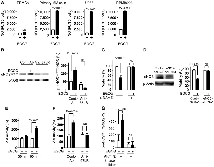
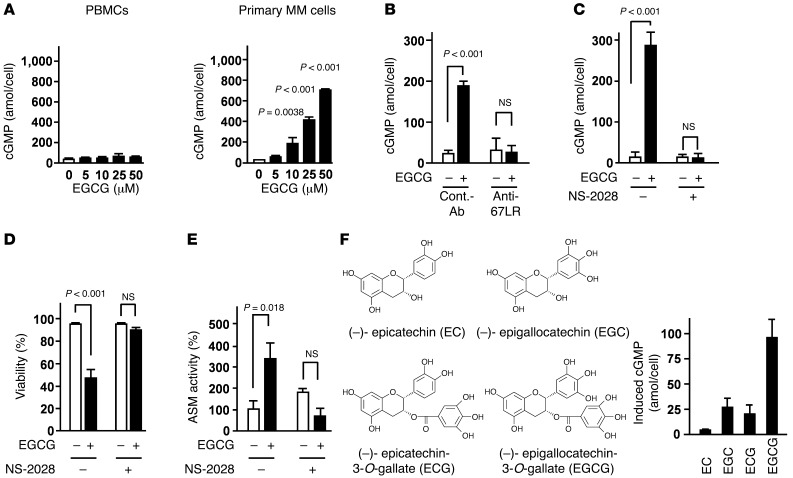
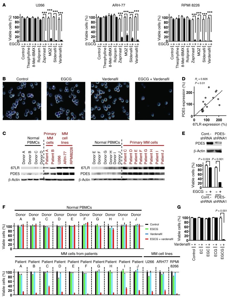
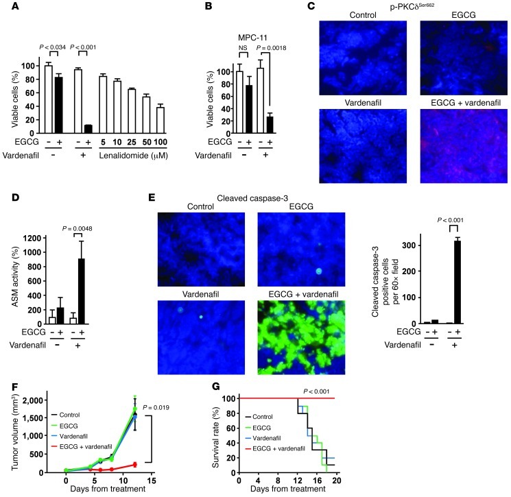
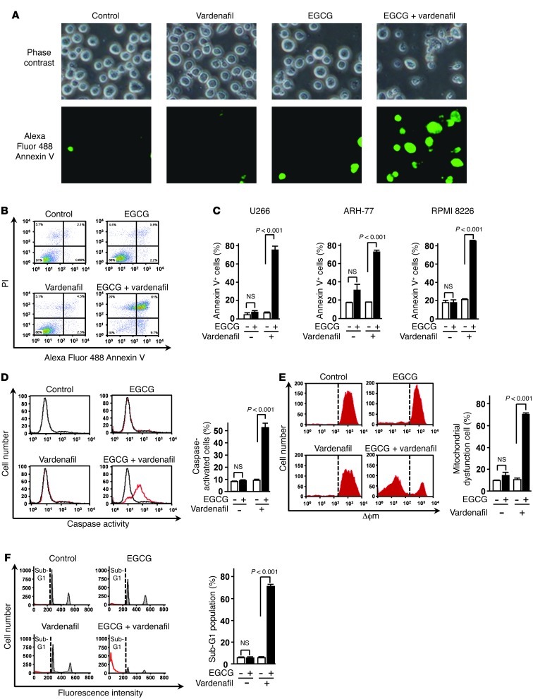
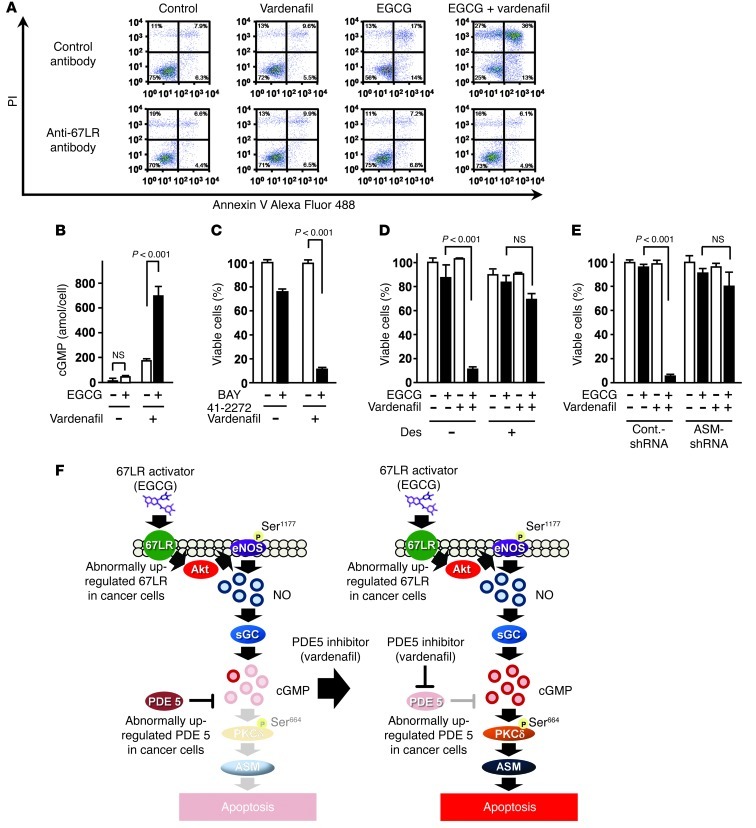
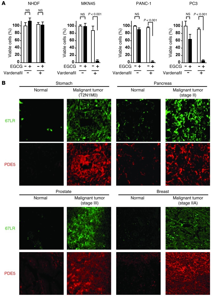
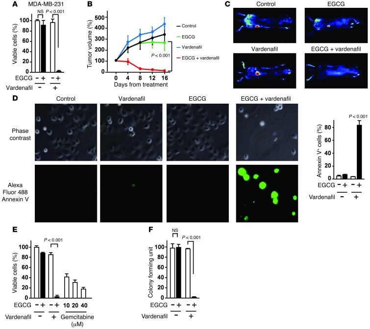
The 67-kDa laminin receptor (67LR) is a laminin-binding protein overexpressed in various types of cancer, including bile duct carcinoma, colorectal carcinoma, cervical cancer, and breast carcinoma. 67LR plays a vital role in growth and metastasis of tumor cells and resistance to chemotherapy. Here, we show that 67LR functions as a cancer-specific death receptor. In this cell death receptor pathway, cGMP initiated cancer-specific cell death by activating the PKCδ/acid sphingomyelinase (PKCδ/ASM) pathway. Furthermore, upregulation of cGMP was a rate-determining process of 67LR-dependent cell death induced by the green tea polyphenol (-)-epigallocatechin-3-O-gallate (EGCG), a natural ligand of 67LR. We found that phosphodiesterase 5 (PDE5), a negative regulator of cGMP, was abnormally expressed in multiple cancers and attenuated 67LR-mediated cell death. Vardenafil, a PDE5 inhibitor that is used to treat erectile dysfunction, significantly potentiated the EGCG-activated 67LR-dependent apoptosis without affecting normal cells and prolonged the survival time in a mouse xenograft model. These results suggest that PDE5 inhibitors could be used to elevate cGMP levels to induce 67LR-mediated, cancer-specific cell death.


Figures

Figure 1
Figure 1. 67LR-dependent NO production by activation of Akt and eNOS is a key event in the EGCG-induced cell death pathway.
(A) Primary MM cells, MM cell lines (U266 and RPMI8226), and normal PBMCs were cultured for 3 hours in the absence or presence of 5 μM EGCG. NO production was measured using the fluorescent probe DAF-2DA. FU, fluorescence units. (B) Cells were pretreated with control antibody or anti-67LR antibody (20 μg/ml) and treated or not with EGCG for 3 hours. Phosphorylation of eNOS at Ser1177 was measured by Western blotting. Lanes were run on the same gel but were noncontiguous (white lines). (C) U266 cells were pretreated or not with the NOS inhibitor l-NAME (10 mM), then cultured in medium with or without 10 μM EGCG for 96 hours. (D) Left: Immunoblot analyses of eNOS knockdown in U266 cells. Right: Sensitivity of U266 cells to EGCG (10 μM for 96 hours) after eNOS knockdown. (E) Effect of 5 μM EGCG on Akt activity. (F) Cells were pretreated with control antibody or anti-67LR antibody (20 μg/ml) and treated or not with 5 μM EGCG for 1 hour. (G) U266 cells were pretreated or not with 5 μM AKT1/2 kinase inhibitor, then cultured in medium with or without 5 μM EGCG for 3 hours. All data are mean ± SEM.
Figure 2
Figure 2. 67LR acts as a death receptor via cancer-specific cGMP upregulation.
(A) Effect of EGCG on cGMP levels in normal PBMCs and primary MM cells for 3 hours. (B) U266 cells were preincubated with anti-67LR antibody (20 μg/ml) or IgM control antibody (20 μg/ml), then treated or not with 10 μM EGCG for 3 hours. (C) Effect of the sGC inhibitor NS-2028 on cGMP upregulation. U266 cells were preincubated or not with 5 μM NS-2028 for 1 hour, then treated or not with 10 μM EGCG for 3 hours. (D) Effect of NS-2028 on cell death induced by EGCG. U266 cells were preincubated or not with 5 μM NS-2028 for 1 hour, then treated or not with 10 μM EGCG for 96 hours. (E) Effect of NS-2028 on EGCG-induced ASM activation, measured by TLC analyses. U266 cells were preincubated or not with 5 μM NS-2028 for 1 hour, then treated or not with 10 μM EGCG for 3 hours. (F) U266 cells were treated with 10 μM EGCG or its analogs for 3 hours, and cGMP levels in cells were measured using a competitive immunoassay. n = 3 per group. All data are mean ± SEM.
Figure 3
Figure 3. Abnormal overexpression of PDE5 attenuates EGCG-induced cell death in MM cells.
(A) MM cells were pretreated with the PDE1 inhibitor 8-Met-IBMX (20 μM), with the PDE4 inhibitor rolipram (10 μM), with the PDE5 inhibitors zaprinast (10 μM), sildenafil (10 μM), vardenafil (5 μM), and MQZ (10 μM), or with theophylline (20 μM), then treated or not with 5 μM EGCG for 96 hours. ***P < 0.001. (B) Cells were treated with or without 5 μM vardenafil and/or 5 μM EGCG for 96 hours. Phase-contrast images were taken by optical microscopy. Original magnification, ×20. (C) Expression of 67LR and PDE5 proteins in patient cells and normal PBMCs, assessed by immunoblotting. Lanes were run on the same gel but were noncontiguous (white lines). (D) Correlation between 67LR expression and PDE5 expression. (E) Top: Immunoblot analyses of PDE5 in U266 cells. Bottom: EGCG sensitivity (5 μM for 96 hours) of U266 cells after PDE5 knockdown. (F) Normal PBMCs from 10 healthy donors, primary MM cells from 10 patients, and MM cell lines were treated with or without 5 μM vardenafil and/or 5 μM EGCG for 96 hours. (G) U266 cells were incubated for 96 hours with or without 5 μM vardenafil and/or 5 μM EGCG analogs (see Figure 2F). n = 3 per group. All data are mean ± SEM.
Figure 4
Figure 4. Effect of EGCG and a PDE5 inhibitor in combination on myeloma cell proliferation in vivo.
(A) Anti-MM effect of EGCG (5 μM) and vardenafil (5 μM) in combination and of lenalidomide for 96 hours in vitro (n = 3). (B) Mouse myeloma MPC-11 cells were pretreated or not with 5 μM vardenafil for 3 hours and cultured in the presence or absence of 5 μM EGCG for 96 hours (n = 3). (CE) MPC-11 cells were injected subcutaneously into female BALB/c mice, and mice (n = 5 per group) were given single i.p. injections of EGCG (15 mg/kg) and/or vardenafil (5 mg/kg). (C) After 6 hours, tumors were excised and evaluated for phosphorylation of PKCδ at Ser662 (corresponding to human p-PKCδSer664). Original magnification, ×60. (D) ASM activation, assessed by TLC analyses. (E) Cleaved caspase-3 was evaluated by immunofluorescence analyses. Original magnification, ×60. (F and G) MPC-11 cells were injected subcutaneously into female BALB/c mice, and mice (n = 10 per group) were given i.p. injections of EGCG (15 mg/kg) and/or vardenafil (5 mg/kg) every 2 days. Statistical analyses of survival curves were undertaken using log-rank analyses of the Kaplan-Meier curves. All data are mean ± SEM.
Figure 5
Figure 5. Caspase activation and collapse of Δϕm are involved in 67LR-dependent cell death.
(A) U266 cells were treated or not for 96 hours with 5 μM EGCG in the presence or absence of 5 μM vardenafil and observed under a fluorescence microscope. Original magnification, ×20. (B and C) Apoptotic cells were double stained with annexin V–Alexa Fluor 488 and PI. (D and E) U266 cells were treated or not for 72 hours with 5 μM EGCG in the presence or absence of 5 μM vardenafil, and caspase activity (D) and collapse of Δϕm (E) were measured. (F) U266 cells were treated for 96 hours with 5 μM EGCG in the presence or absence of 5 μM vardenafil. Flow cytometric analyses of PI staining for cellular DNA content. Sub-G1 fractions were determined by FlowJo. n = 3 per group. All data are mean ± SEM.
Figure 6
Figure 6. Inhibition of PDE5 potentiates 67LR-dependent cell death by enhancing the 67LR-dependent signaling pathway in MM cells.
(A) U266 cells were preincubated for 3 hours with anti-67LR antibody or IgM control antibody, then treated or not for 96 hours with 5 μM EGCG and/or vardenafil. Apoptotic cells were double stained with annexin V–Alexa Fluor 488 and PI and analyzed by flow cytometry. (B) Cells were pretreated or not with 5 μM vardenafil and cultured for 3 hours in the presence or absence of 5 μM EGCG, followed by measurement of the amount of cGMP in the cells. (C) U266 cells were treated or not for 96 hours with the sGC activator BAY 41-2272 (1 μM) in the presence or absence of 5 μM vardenafil. (D) U266 cells were preincubated or not for 3 hours with the ASM-specific inhibitor desipramine (Des; 5 μM), then treated or not for 96 hours with 5 μM EGCG and/or vardenafil. (E) Sensitivity of U266 cells to EGCG and/or vardenafil (5 μM for 96 hours) after ASM knockdown. n = 3 per group. All data are mean ± SEM. (F) The combination of EGCG as a cancer-specific cGMP inducer with an inhibitor targeting cancer-overexpressed PDE5 could be a useful strategy for cancer-selective chemotherapy. Left: In the absence of PDE5 inhibitor, overexpressed PDE5 in cancer attenuates the anticancer effect of EGCG. Right: PDE5 inhibition potentiates the anticancer effect of EGCG.
Figure 7
Figure 7. PDE5 is overexpressed in various types of human cancer.
(A) NHDFs from healthy donors as well as cell lines MKN45 (gastric cancer), PANC-1 (pancreatic cancer), and PC3 (prostate cancer) were cultured for 96 hours with 5 μM vardenafil and/or 5 μM EGCG. (B) Representative images of paraffin sections of various types of tissue from cancer patients. Original magnification, ×60. n = 3 per group for all in vitro studies. All data are mean ± SEM.
Figure 8
Figure 8. PDE5 inhibition potentiates the anticancer effect of EGCG.
(A) MDA-MB-231-RFP cells were cultured for 96 hours with 5 μM vardenafil and/or 5 μM EGCG. (B and C) MDA-MB-231-RFP cells were injected subcutaneously into female nude mice, and mice (n = 8 per group) were given EGCG (15 mg/kg i.p.) and/or vardenafil (5 mg/kg i.p.). (D) PANC-1 cells were treated for 96 hours with 5 μM EGCG and/or 5 μM vardenafil. Cells were treated with annexin V–Alexa Fluor 488. Original magnification, ×20. (E) PANC-1 cells were treated with 5 μM EGCG and/or 5 μM vardenafil or with the conventional drug gemcitabine for 96 hours. (F) Clonogenic assay in PANC-1 cells. 1,500 PANC-1 cells were cultured for 10 days with 5 μM vardenafil in the presence or absence of 5 μM EGCG. n = 3 per group for all in vitro studies. All data are mean ± SEM.

Comment in

Similar articles

Cited by

References

    1. Carroll M, et al. CGP 57148, a tyrosine kinase inhibitor, inhibits the growth of cells expressing BCR-ABL, TEL-ABL, and TEL-PDGFR fusion proteins. Blood. 1997;90(12):4947–4952. - PubMed
    1. Goldenberg MM. Trastuzumab, a recombinant DNA-derived humanized monoclonal antibody, a novel agent for the treatment of metastatic breast cancer. Clin Ther. 1999;21(2):309–318. doi: 10.1016/S0149-2918(00)88288-0. - DOI - PubMed
    1. de Thé H, Lavau C, Marchio A, Chomienne C, Degos L, Dejean A. The PML-RAR alpha fusion mRNA generated by the t(15;17) translocation in acute promyelocytic leukemia encodes a functionally altered RAR. Cell. 1991;66(4):675–684. doi: 10.1016/0092-8674(91)90113-D. - DOI - PubMed
    1. Li D, et al. 67-kDa laminin receptor in human bile duct carcinoma. Eur Surg Res. 2009;42(3):168–173. - PubMed
    1. Sanjuan X, et al. Overexpression of the 67-kD laminin receptor correlates with tumor progression in human colorectal carcinoma. J Pathol. 1996;179(4):376–380. doi: 10.1002/(SICI)1096-9896(199608)179:4<376::AID-PATH591>3.0.CO;2-V. - DOI - PubMed

Publication types

MeSH terms