. 2013 Feb;123(2):874-86.
doi: 10.1172/JCI63324. Epub 2013 Jan 25.

Behavioral stress accelerates prostate cancer development in mice

Affiliations

Behavioral stress accelerates prostate cancer development in mice

Sazzad Hassan et al. J Clin Invest. 2013 Feb.

Abstract

Prostate cancer patients have increased levels of stress and anxiety. Conversely, men who take beta blockers, which interfere with signaling from the stress hormones adrenaline and noradrenaline, have a lower incidence of prostate cancer; however, the mechanisms underlying stress-prostate cancer interactions are unknown. Here, we report that stress promotes prostate carcinogenesis in mice in an adrenaline-dependent manner. Behavioral stress inhibited apoptosis and delayed prostate tumor involution both in phosphatase and tensin homolog-deficient (PTEN-deficient) prostate cancer xenografts treated with PI3K inhibitor and in prostate tumors of mice with prostate-restricted expression of c-MYC (Hi-Myc mice) subjected to androgen ablation therapy with bicalutamide. Additionally, stress accelerated prostate cancer development in Hi-Myc mice. The effects of stress were prevented by treatment with the selective β2-adrenergic receptor (ADRB2) antagonist ICI118,551 or by inducible expression of PKA inhibitor (PKI) or of BCL2-associated death promoter (BAD) with a mutated PKA phosphorylation site (BADS112A) in xenograft tumors. Effects of stress were also blocked in Hi-Myc mice expressing phosphorylation-deficient BAD (BAD3SA). These results demonstrate interactions between prostate tumors and the psychosocial environment mediated by activation of an adrenaline/ADRB2/PKA/BAD antiapoptotic signaling pathway. Our findings could be used to identify prostate cancer patients who could benefit from stress reduction or from pharmacological inhibition of stress-induced signaling.


Figures
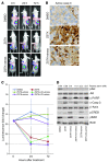
Figure 1
Figure 1. Stress or adrenaline prevents apoptosis induced by PI3K inhibitor in prostate cancer xenografts.
(A) Representative images of mice with C42LucBAD xenografts. Mice were injected with DMSO, ZSTK474 (ZSTK), or ZSTK474 followed by immobilization stress. Luminescence of xenograft tumors was measured before injection of PI3K inhibitors (0 hour) and after injections at 24 and 72 hours. (B) Immunohistochemical analysis of cleaved caspase-3 in C42LucBAD xenograft tumors. Mice were treated as in A, and xenografts were excised 6 hours after injections. Scale bars: 50 μm. (C) Dynamics of luminescence in C42LucBAD xenograft tumors. Mice were injected with DMSO (n = 3), ZSTK474 (n = 3), ZSTK474 followed by immobilization stress (n = 3), ZSTK474 and adrenaline (adren; n = 3), ZSTK474 and ICI118,551 (ICI) followed by immobilization stress (n = 3), or ZSTK474, ICI118,551, and adrenaline (n = 4). Error bars show SD from the average of measurements in at least 3 mice. Comparisons between pairs of groups were performed using t tests derived from the overall ANOVA model. Significant between-group differences over time were as follows: P < 0.02, ZSTK vs. DMSO; P < 0.01, ZSTK+stress vs. ZSTK; P < 0.03, ZSTK+adren vs. ZSTK; P < 0.003, ZSTK+ICI+stress vs. ZSTK+stress; P < 0.006, ZSTK+ICI+adren vs. ZSTK+adren. (D) Western blot analysis of C42LucBAD xenograft tumor tissues excised 6 hours after injection of ZSTK474 and/or ICI118,551 (ICI) into mice that were then subjected to immobilization stress, left intact, or injected with epinephrine. At the time of xenograft excision, blood was collected for adrenaline measurements (shown above blots).
Figure 2
Figure 2. Activation of PKA is necessary for stress- or adrenaline-induced protection from apoptosis in prostate cancer xenografts.
(A) Analysis of C42LucPKI xenograft tumors by Western blotting. ZSTK474 inhibited pAktS473 and pBADS112 and induced cleavage of PARP and caspase-3. Stress or adrenaline induced pBADS112 and pCREBS133 and inhibited cleavage of PARP and caspase-3. These effects of stress or adrenaline were blocked by doxycycline-induced (Dox) PKI-GFP expression. At the time of tumor excision, blood was collected for adrenaline measurements (shown above blots). (B and C) Effects of stress and adrenaline on tumor luminescence depend on PKA activity. Mice were injected with DMSO as a control or with ZSTK474, with or without adrenaline or subsequent immobilization stress. Comparisons between groups were performed using t tests derived from the overall ANOVA model. (B) In mice that did not receive doxycycline, significant differences in luminescence across time were found in ZSTK+stress (P < 0.0002) and ZSTK+adren (P < 0.0002) groups versus ZSTK. (C) These differences were eliminated by doxycycline-induced PKI-GFP expression (P > 0.63 and P > 0.13, respectively). Error bars in B and C show the SD from the average of measurements in at least 4 mice.
Figure 3
Figure 3. Activation of pBADS112 is necessary for stress- or adrenaline-induced protection from apoptosis in prostate cancer xenografts.
(A) Analysis of C42LucBAD1SA xenograft tumors by Western blotting. ZSTK474 inhibited pAktS473 and pBADS112 and induced cleavage of PARP and caspase-3. Stress or adrenaline induced pBADS112 and pCREBS133 and inhibited cleavage of PARP and caspase-3. Effects of stress or adrenaline on apoptosis (cleavage of caspase-3 and PARP) were blocked by doxycycline-induced expression of mutant HA-BADS112A. Arrowheads denote HA-BAD1SA (top) and endogenous BAD (bottom). The inset at right from a pBAD blot shows lysates of C42LucBAD cells that served as positive control for phosphorylated HA-BAD. Mutant HA-BADS112A (lanes 5–7) could not be phosphorylated and was not recognized by pBADS112-specific antibodies. (B and C) Effects of stress or adrenaline on tumor luminescence depend on pBADS112. (B) In mice that did not receive doxycycline, luminescence in ZSTK+stress and ZSTK+adren groups was highly significantly different compared with the ZSTK group (P < 0.0001 for both). (C) These differences were completely eliminated by doxycycline-induced expression of pBADS112-deficient HA-BAD1SA (P > 0.65 and P > 0.52, respectively). Error bars in B and C show the SD from the average of measurements in at least 4 mice.
Figure 4
Figure 4. Stress induces BAD phosphorylation and inhibits cleavage of PARP and caspase-3 in Hi-Myc mouse prostate glands.
WT and Hi-Myc mice were subjected to recurrent 1-hour immobilization stress at 12-hour intervals for 7 consecutive days; blood and prostates were collected immediately after the last stress procedure. ICI118,551 was given 30 minutes before stress. (A) Western blot analysis of DLP glands excised from intact (denoted “calm” or “C”) and stressed (“stress” or “S”) mice was conducted with antibodies to pCREBS133, pBADS112, cleaved caspase-3, cleaved PARP, α-tubulin, and c-Myc. 4 representative samples from each treatment group are shown. (B) Densitometric analysis of Western blots revealed statistically significant increases of BAD and CREB phosphorylation in stressed versus intact mice (pBADS112, P = 0.005 between WT groups; P = 0.02 between Hi-Myc groups; pCREBS133, P = 0.02 between WT groups; P = 0.0019 between Hi-Myc groups) and reduced cleavage of caspase-3 (P = 0.005) and PARP (P = 0.01) in stressed versus intact Hi-Myc mice. These effects of stress were completely eliminated by ICI118,551. Each experimental group contained at least 5 mice. Error bars represent SD from the average of at least 5 samples.
Figure 5
Figure 5. Stress accelerates PIN growth and inhibits apoptosis in DLP glands of Hi-Myc mice.
(A) Stress increased prostate weight. Mouse prostates (AP, DLP, and VP lobes) were dissected and weighed, and the total prostate wet weight was expressed as mg/25 g body weight. Statistically significant differences were observed between intact and stressed Hi-Myc mice (P = 0.002), but not between WT groups (P = 1.00). This difference in Hi-Myc mice was eliminated by ICI118,551 injection prior to stress (P = 0.49). (B) Stress reduced apoptosis in DLP glands. Percent cleaved caspase-3–labeled cells in immunostained sections was determined relative to the total number of glandular epithelial cells in whole sections of DLP. Representative images of cleaved caspase-3 IHC-stained sections from DLP of intact or stressed Hi-Myc mice are also shown. Scale bars: 50 μm. Insets show the original images (×40 objective) enlarged ×5. (C) Stress increased PIN in DLP glands of Hi-Myc mice. Percent PIN area was determined as area of PIN divided by total DLP glandular area. Representative microphotographs of H&E-stained sections from DLP glands of intact and stressed Hi-Myc mice are also shown. Scale bars: 50 μm. See Supplemental Figure 8D for morphology of mouse PIN. Error bars in AC show SD from the average of at least 5 samples.
Figure 6
Figure 6. Antiapoptotic and tumor-promoting effects of stress are eliminated in BAD3SA/WT knockin mice.
(A) Western blot analysis of DLP glands excised from 12-week-old intact and stressed Hi-MycBAD3SA/WT mice was conducted with antibodies to pCREBS133, pBADS112, cleaved caspase-3, cleaved PARP, and β-actin. (B) Densitometric analysis of Western blots revealed statistically significant increases of pCREBS133 in stressed versus intact Hi-MycBAD3SA/WT mice (P = 0.003); however, no significant differences between intact and stressed mice were found in pBADS112 (P = 0.29), cleaved caspase-3 (P = 0.47), or cleaved PARP (P = 0.25). Lysates of 4 intact mice were used for densitometry (2 mice with adrenalin levels greater than 1 nM were excluded as erroneously stressed); the stressed group contained 6 mice. (C) Prostates of intact and stressed Hi-MycBAD3SA/WT (n = 5 per group) and WTBAD3SA/WT (n = 3 per group) mice were analyzed as in Figure 3B. (D) Microphotographs of dissected prostate glands from Hi-MycBAD3SA/WT or WTBAD3SA/WT mice either subjected to repeated immobilization stress for 7 days or left intact. (E) Prostates of intact and stressed Hi-MycBAD3SA/WT mice were analyzed as in Figure 5B (n = 5 per group). Bars in B, C, and E show SD. (F) Representative images of H&E-stained sections of DLP glands of intact or stressed Hi-MycBAD3SA/WT mice. Scale bars: 50 μm.
Figure 7
Figure 7. Stress delays bicalutamide-induced involution and apoptosis in Hi-Myc prostates via the ADRB2/BAD pathway.
Hi-Myc mice were subjected to subcutaneous injection of bicalutamide (bicalut.; 50 mg/kg, once daily) and recurrent 1-hour immobilization stress at 12-hour intervals for 3 consecutive days; blood and prostates were collected immediately after the last stress procedure. ICI118,551 was given 30 minutes before stress. (A) Stress delayed bicalutamide-induced prostate involution in Hi-Myc mice. Mouse prostates (AP, DLP, and VP lobes) were dissected and weighed, and the total prostate wet weight was expressed as mg/25 g body weight. Statistically significant differences were observed between intact and bicalutamide-treated intact Hi-Myc mice (P = 0.01) and between bicalutamide-treated intact and stressed Hi-Myc mice (P = 0.002). The effect of bicalutamide on prostate weight was completely eliminated with ICI118,551 (P = 0.54) and significantly reduced from 2- to 0.2-fold in Hi-MycBAD3SA/WT mice (P = 0.049). Representative images of prostates of Hi-Myc intact and stressed mice treated with bicalutamide are also shown. (B) Stress delayed bicalutamide-induced apoptosis in DLP glands of Hi-Myc mice (P = 0.002). The effect of stress on bicalutamide-induced apoptosis was eliminated in Hi-Myc mice injected with ICI118,551 (P = 0.84) and in compound transgenic Hi-MycBAD3SA/WT mice (P = 0.47). Representative images of cleaved caspase-3 IHC-stained sections from DLP of intact and stressed Hi-Myc mice treated with bicalutamide are also shown. Scale bars: 50 μm. Insets show the original images (×40 objective) enlarged ×2.33.
Figure 8
Figure 8. Stress inhibits bicalutamide-induced cleavage of PARP and caspase-3 through ADRB2 and BAD phosphorylation in prostates of Hi-Myc mice.
(A) Western blot analysis of prostate glands excised from intact mice and bicalutamide-injected intact or stressed mice was conducted with antibodies to pCREBS133, pBADS112, cleaved caspase-3, cleaved PARP, β-actin, and probasin. 3 representative samples for each treatment group are shown. (B) Densitometric analysis of Western blots showed significant increases of BAD and CREB phosphorylation in stressed versus intact Hi-Myc mice (pCREBS133, P = 0.01; pBADS112, P = 0.008), which was blocked by ICI118,551. No significant differences were found in pCREBS133 (P = 0.35) or pBADS112 (P = 0.34) between the intact and stressed bicalutamide treatment groups. In Hi-MycBAD3SA/WT mice, stress significantly increased pCREBS133 (P = 0.003), but not pBADS112 (P = 0.15). (C) Stress decreased cleavage of caspase-3 and PARP induced by bicalutamide (P = 0.003, intact vs. bicalutamide intact; P = 0.004, bicalutamide intact vs. bicalutamide stress). These effects were eliminated in mice treated with ICI118,551 and in Hi-MycBAD3SA/WT mice (P > 0.5). P values were similar for cleaved caspase-3 and PARP. (D) Bicalutamide treatment significantly decreased probasin expression (P = 0.01), and stress did not affect probasin expression in prostates of bicalutamide-treated mice (P = 0.58). Each experimental group contained at least 5 mice. Error bars in BD show SD from the average of at least 4 samples.
Figure 9
Figure 9. A vicious circle of stress signaling in prostate cancer.
Psychoemotional stress activates the hypothalamic-pituitary-adrenal axis (HPA); as a result, blood levels of adrenaline increase and activate ADRB2/PKA/BAD antiapoptotic signaling in prostate cells. Constitutive activation of this signaling pathway in stressed patients (Supplemental Figure 12) may trigger a vicious circle: stress and anxiety from a diagnosis of prostate cancer increase adrenaline levels that in turn reduce efficacy of anticancer treatments.

Comment in

  • Why stress is BAD for cancer patients.
    Nagaraja AS, Armaiz-Pena GN, Lutgendorf SK, Sood AK. Nagaraja AS, et al. J Clin Invest. 2013 Feb;123(2):558-60. doi: 10.1172/JCI67887. Epub 2013 Jan 25. J Clin Invest. 2013. PMID: 23348736 Free PMC article.

References

    1. Berkow SE, Barnard ND, Saxe GA, Ankerberg-Nobis T. Diet and survival after prostate cancer diagnosis. Nutr Rev. 2007;65(9):391–403. doi: 10.1111/j.1753-4887.2007.tb00317.x. - DOI - PubMed
    1. Saxe GA, Major JM, Nguyen JY, Freeman KM, Downs TM, Salem CE. Potential attenuation of disease progression in recurrent prostate cancer with plant-based diet and stress reduction. Integr Cancer Ther. 2006;5(3):206–213. doi: 10.1177/1534735406292042. - DOI - PubMed
    1. Ornish D, et al. Changes in prostate gene expression in men undergoing an intensive nutrition and lifestyle intervention. Proc Natl Acad Sci U S A. 2008;105(24):8369–8374. doi: 10.1073/pnas.0803080105. - DOI - PMC - PubMed
    1. Spiegel D, Giese-Davis J. Depression and cancer: mechanisms and disease progression. Biol Psychiatry. 2003;54(3):269–282. doi: 10.1016/S0006-3223(03)00566-3. - DOI - PubMed
    1. Armaiz-Pena GN, Lutgendorf SK, Cole SW, Sood AK. Neuroendocrine modulation of cancer progression. Brain Behav Immun. 2009;23(1):10–15. doi: 10.1016/j.bbi.2008.06.007. - DOI - PMC - PubMed

Publication types

MeSH terms