. 2013 Jan 15;27(2):163-8.
doi: 10.1101/gad.207043.112.

Centriole distal appendages promote membrane docking, leading to cilia initiation

Affiliations

Centriole distal appendages promote membrane docking, leading to cilia initiation

Barbara E Tanos et al. Genes Dev. .

Abstract

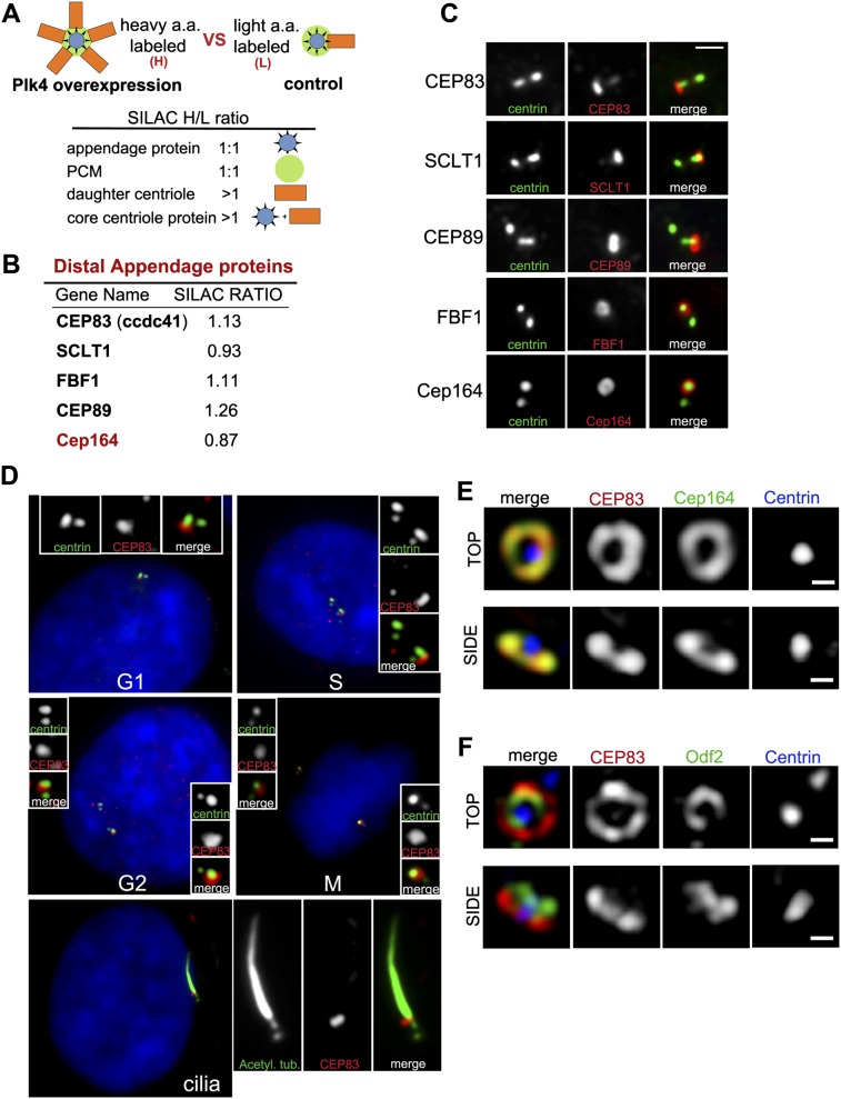
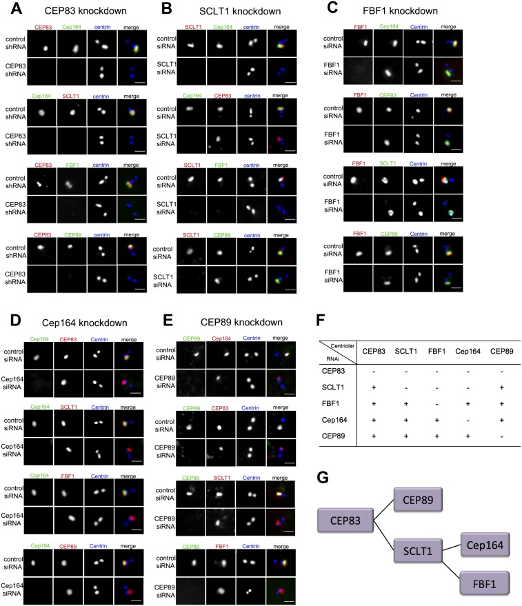
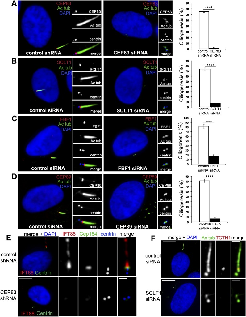
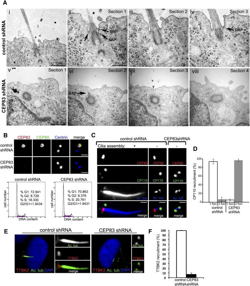
The distal appendages (DAPs) of centrioles have been proposed to anchor cilia to the plasma membrane, but their molecular composition, assembly, and exact function in ciliogenesis remain poorly understood. Using quantitative centrosome proteomics and superresolution microscopy, we identified five DAP components, including one previously described (CEP164), one partially characterized (CEP89 [ccdc123]), and three novel (CEP83 [ccdc41], SCLT1, and FBF1) DAP proteins. Analyses of DAP assembly revealed a hierarchy. CEP83 recruits both SCLT1 and CEP89 to centrioles. Subsequent recruitment of FBF1 and CEP164 is independent of CEP89 but mediated by SCLT1. All five DAP components are essential for ciliogenesis; loss of CEP83 specifically blocks centriole-to-membrane docking. Undocked centrioles fail to recruit TTBK2 or release CP110, the two earliest modifications found on centrioles prior to cilia assembly, revealing centriole-to-membrane docking as a temporal and spatial cue promoting cilia initiation.


Figures

Figure 1.
Figure 1.
Identification of novel DAP proteins. (A) Schematic of SILAC experiment. DAP proteins belong to the group of proteins showing a 1:1 ratio. (B) Summary of the screen for DAP proteins. The SILAC ratio (H/L) is indicated. (C) RPE-1 cells in G1 were stained with antibodies to the indicated DAP proteins (red) or to centrin (green). Note the asymmetrical localization of DAP proteins. Bar, 1 μm. (D) Subcellular distribution of CEP83. RPE-1 cells stably expressing GFP-centrin 2 (green) were stained with an antibody to CEP83 (red) and DAPI (blue). Cell cycle stages are indicated; note that CEP83 is incorporated into centrioles during G2. (Bottom panel) In ciliated cells where primary cilia are marked with antibodies against acetylated α-tubulin (green), CEP83 localizes to the ciliary base. (E,F) RPE-1 cells stably expressing HA-tagged CEP83 were stained with antibodies to HA (red), Cep164 (green) (E) or Odf2 (green) (F) and centrin (blue) and examined with an OMX superresolution microscope (Applied Precision, Inc.). Note that CEP83 colocalizes with the DAP protein Cep164 both in top view (top panels) and side view (bottom panels) but does not colocalize with the SAP protein Odf2. Small bar, 200 nm.
Figure 2.
Figure 2.
The DAP assembly pathway. (A) RPE1 cells were retrovirally transduced with control shRNA or a CEP83 targeting shRNA as indicated and stained with antibodies to CEP83 (red) and to the other DAPs identified (green) (see Fig. 1A). Centrioles are marked with centrin (blue). (First panel) Note that CEP83 is required for the centriolar localization of the known DAP protein Cep164 as well as all of the other DAP components. The second panel from the top shows that SCLT1 is absent from CEP83-depleted centrioles marked by the absence of Cep164. Bar, 1 μm. (B) Cells transfected with control siRNA or SCLT1 siRNA were stained with antibodies to SCLT1 (red) and to the other DAP proteins identified (green). Centrioles are marked with centrin (blue). Note that Cep164 is absent from SCLT1-depleted centrioles (first panel), whereas CEP83 is still present (second panel). Bar, 1 μm. (C) Cells transfected with control siRNA or FBF1 siRNA were stained with antibodies to FBF1 (red) and to all other DAP components (green). Centrioles are marked with centrin (blue). Bar, 1 μm. (D) Cells transfected with control siRNA or Cep164 siRNA were stained with antibodies to Cep164 (green) and to the other DAP proteins (red). Centrioles are marked with centrin (blue). Bar, 1 μm. (E) Cells transfected with control siRNA or CEP89 siRNA were stained with antibodies to CEP89 (green) and to the other DAP proteins (red). Centrioles are marked with centrin (blue). Bar, 1 μm. (F) A table showing the interdependency of centriolar localization for the proteins identified. (G) Assembly pathway of centriolar DAPs.
Figure 3.
Figure 3.
All DAP components are required for ciliogenesis. (A) RPE-1 cells stably expressing centrin-GFP (blue) were retrovirally transduced with control shRNA (left panel) or CEP83 shRNA (right panel), grown in serum-free medium for 48 h, and then fixed and stained with antibodies to CEP83 (red) and acetylated tubulin (green). DNA staining with DAPI is shown in blue. A quantification of this result is shown in the graph on the right (>300 cells). (B–D) RPE-1 cells stably expressing centrin-GFP (blue) were transfected with control siRNA (left panels) or SCLT1 (B), FBF1 (C), or CEP89 siRNA (D) (right panels), grown in serum-free medium for 48 h, and then fixed and stained with antibodies to SCLT1 (B), FBF1 (C), or CEP89 (red) and acetylated tubulin (green). DNA staining with DAPI is shown in blue. A graph with the quantification of each result is shown (>300 cells). (E) RPE-1 cells retrovirally transduced with control shRNA (top panels) or CEP83 shRNA (bottom panels) and stained with antibodies against IFT88 (red) or Cep164 (green) and centrin (blue). Note that in RPE-1 control cells, IFT88 localizes to both the basal bodies and ciliary axoneme. These localizations are lost in CEP83-depleted cells (marked by the absence of Cep164). (F) RPE-1 cells were transfected with control siRNA (top panels) or SCLT1 siRNA (bottom panels) and stained with antibodies to acetylated tubulin (green) or TCTN1 (red). DNA staining with DAPI is shown in blue. Large bar, 5 μm; small bar, 1 μm.
Figure 4.
Figure 4.
Coordination of centriole-to-membrane docking and cilia initiation. (A) IMCD3 cells were retrovirally transduced with either a luciferase control shRNA (panels i–iv) or a CEP83 targeting shRNA (panels v–viii). Three days after infection, cells were plated on top of polystyrene transwell filters and allowed to polarize and form a monolayer for 72 h. Filters were then fixed and processed for transmission EM. (Panel i) Thin section showing a centriole pair and cilia in IMCD3 control cells (25K). Bar, 1 μm. (Panel ii) Centriole pair in panel i at a higher magnification (50K). Note the mother centriole docked to the plasma membrane and daughter centriole in cross-section. DAPs and SAPs can be clearly seen (arrows, indicated). Bar, 500 nm. (Panel iii) Consecutive section showing the same centriole pair as in panel ii and the beginning of the ciliary axoneme. (Panel iv) Consecutive section showing the same centriole pair as in panel iii. Note that the tip of a distal appendage points to the site of attachment (arrow). (Panel v) Mother centriole of a pair in an IMCD3 cell depleted of CEP83 (50K). Bar, 500 nm. (Panel vi) Consecutive section of centriole in panel v. Note that the centriole is close to the apical cortex but has not docked; one subdistal appendage can be seen (arrow). (Panel vii) Continuous section. (Panel viii) Continuous section showing the other centriole of the pair. (B) RPE-1 cells stably expressing centrin-GFP (blue) were retrovirally transduced with control shRNA (top panels) or CEP83 shRNA (bottom panels), serum-starved for 48 h, and stained with antibodies to CEP83 and CEP89. Note that in RPE-1 cells, CEP83 is required for CEP89 localization to centrioles. Bar, 1 μm. The bottom panels show the cell cycle profile of control RPE-1 cells (left) and CEP83-depleted cells (right). (C) RPE-1 cells transduced with control shRNA or CEP83 shRNA were serum-starved for 48 h, fixed, and stained for CEP89 (red), CP110 (green), and acetylated tubulin (blue). Note that CP110 removal normally occurs at mature centrioles before cilia assembly initiates but fails in CEP83-depleted centrioles. Bar, 1 μm. (D) Quantification of CP110 recruitment. (E) RPE-1 cells transduced with control shRNA or CEP83 shRNA were serum-depleted for 48 h, fixed, and stained for TTKB2 (red) and acetylated tubulin (green). Note that in control cells, TTKB2 localizes to the centriole that supports cilia formation, whereas in CEP83-depleted cells, the recruitment of TTKB2 fails. (F) Quantification of TTBK2 recruitment.

Similar articles

Cited by

References

    1. Anderson RG 1972. The three-dimensional structure of the basal body from the rhesus monkey oviduct. J Cell Biol 54: 246–265 - PMC - PubMed
    1. Chih B, Liu P, Chinn Y, Chalouni C, Komuves LG, Hass PE, Sandoval W, Peterson AS 2012. A ciliopathy complex at the transition zone protects the cilia as a privileged membrane domain. Nat Cell Biol 14: 61–72 - PubMed
    1. Eggenschwiler JT, Anderson KV 2007. Cilia and developmental signaling. Annu Rev Cell Dev Biol 23: 345–373 - PMC - PubMed
    1. Goetz SC, Liem KF Jr, Anderson KV 2012. The spinocerebellar ataxia-associated gene tau tubulin kinase 2 controls the initiation of ciliogenesis. Cell 151: 847–858 - PMC - PubMed
    1. Graser S, Stierhof YD, Lavoie SB, Gassner OS, Lamla S, Le Clech M, Nigg EA 2007. Cep164, a novel centriole appendage protein required for primary cilium formation. J Cell Biol 179: 321–330 - PMC - PubMed

Publication types