. 2013 Mar 1;187(5):509-17.
doi: 10.1164/rccm.201211-1983OC. Epub 2013 Jan 24.

Mechanisms of cardiac and renal dysfunction in patients dying of sepsis

Affiliations

Mechanisms of cardiac and renal dysfunction in patients dying of sepsis

Osamu Takasu et al. Am J Respir Crit Care Med. .

Abstract

Rationale: The mechanistic basis for cardiac and renal dysfunction in sepsis is unknown. In particular, the degree and type of cell death is undefined.

Objectives: To evaluate the degree of sepsis-induced cardiomyocyte and renal tubular cell injury and death.

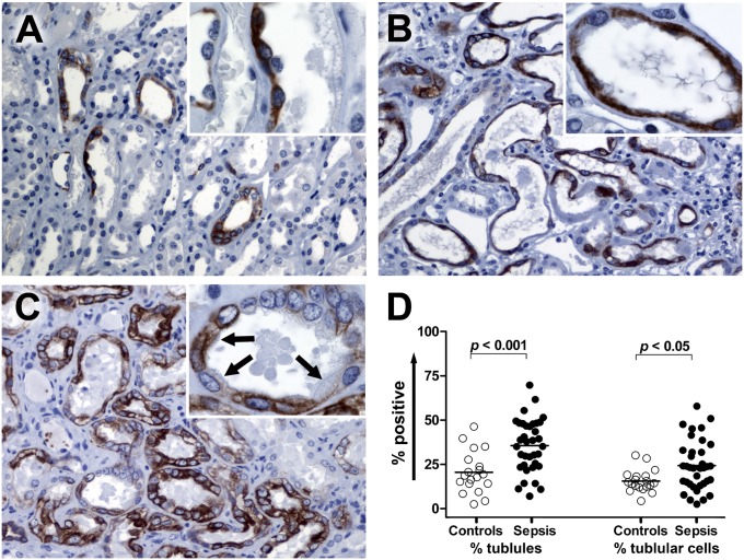
Methods: Light and electron microscopy and immunohistochemical staining for markers of cellular injury and stress, including connexin-43 and kidney-injury-molecule-1 (Kim-1), were used in this study.

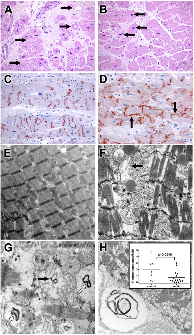
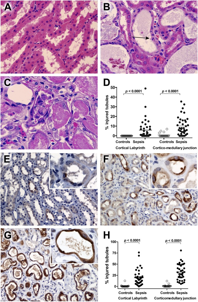
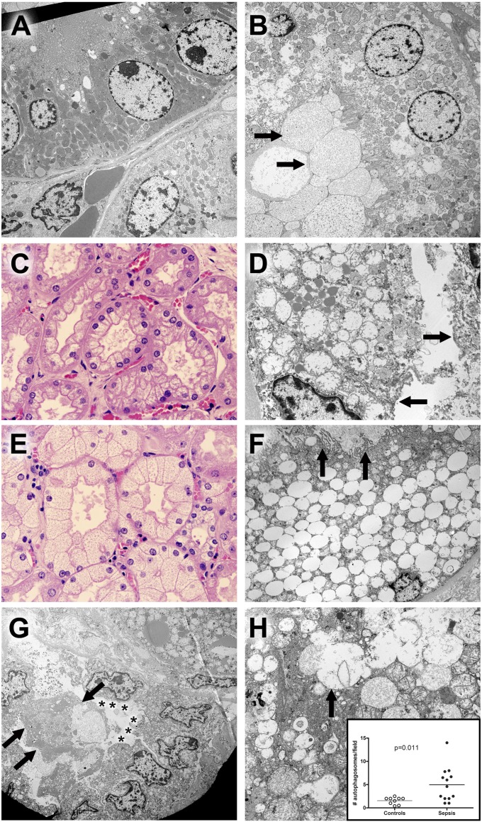
Measurements and main results: Rapid postmortem cardiac and renal harvest was performed in 44 septic patients. Control hearts were obtained from 12 transplant and 13 brain-dead patients. Control kidneys were obtained from 20 trauma patients and eight patients with cancer. Immunohistochemistry demonstrated low levels of apoptotic cardiomyocytes (<1-2 cells per thousand) in septic and control subjects and revealed redistribution of connexin-43 to lateral membranes in sepsis (P < 0.020). Electron microscopy showed hydropic mitochondria only in septic specimens, whereas mitochondrial membrane injury and autophagolysosomes were present equally in control and septic specimens. Control kidneys appeared relatively normal by light microscopy; 3 of 20 specimens showed focal injury in approximately 1% of renal cortical tubules. Conversely, focal acute tubular injury was present in 78% of septic kidneys, occurring in 10.3 ± 9.5% and 32.3 ± 17.8% of corticomedullary-junction tubules by conventional light microscopy and Kim-1 immunostains, respectively (P < 0.01). Electron microscopy revealed increased tubular injury in sepsis, including hydropic mitochondria and increased autophagosomes.

Conclusions: Cell death is rare in sepsis-induced cardiac dysfunction, but cardiomyocyte injury occurs. Renal tubular injury is common in sepsis but presents focally; most renal tubular cells appear normal. The degree of cell injury and death does not account for severity of sepsis-induced organ dysfunction.


Figures

Figure 1.
Figure 1.
Light microscopy and ultrastructural findings in heart. (A) Myocytolysis is common in control cardiac myocytes. Hearts were obtained from septic or control patients. Control hearts were failing hearts from patients undergoing heart transplantation or hearts from brain-dead organ donors whose hearts were not acceptable for transplantation (see Methods). Although most myocardial cells were unremarkable in most histologic fields in control and septic samples, large, pale vacuoles in subendocardial myocytes (myocytolysis; arrows), a putative feature of reversible hypoxic injury, were more common in control samples (P < 0.01) (Figure E1). Note also expansion of interstitial fibrous tissue and enlargement of myocyte nuclei (hypertrophy). Original magnification: ×200. (B) Myocyte hypertrophy is common in septic cardiac samples. Although not specific for sepsis, many septic hearts exhibited conspicuous enlargement of myocyte nuclei (hypertrophy; arrows), often in a setting in interstitial fibrosis. Original magnification: ×200. (C) Connexin-43 is present on intercalated discs in control hearts. Strong granular staining of the intercalated discs is seen in this example. Punctate staining of cytoplasm away from the intercalated discs most likely represents intracellular microtubule-mediated transport. Original magnification: ×200. (D) Connexin-43 localizes to lateral cell membranes in some septic specimens. In addition to labeling the intercalated discs, antibodies to connexin-43 also reveal reactivity along lateral myocyte membranes (arrows) in a subset of septic samples. Lateralization of connexin-43 was more common in septic patients (P < 0.05) (Fig. E1). Original magnification: ×400. (E) Electron microscopy of control cardiac muscle. Contractile elements, with distinct Z-bands, are aligned in register. Mitochondria and other organelles occupy space between myofilaments and contractile fibers; most appear normal. Elements of sarcomplasmic reticulum (T tubules) are also evident. A portion of a normal-appearing intercalated disc is present (open arrow). Isolated lipid droplets are present. Original magnification: ×9,730. (F) Electron microscopy of septic cardiac muscle. Contractile elements are in register but are mildly splayed in areas. Mitochondria are relatively normal in appearance but with focal hydropic change (arrow); crista are generally intact. Lipid is present. Original magnification: ×12,800. (G) Mitochondrial abnormalities in sepsis. Hydropic change (edema of the mitochondrial matrix) is associated with cystic alterations of the crista (closed arrow) and collapse into small myelin-like clusters (open arrow). Original magnification: ×18,400. (H) Autophagy in cardiac muscle. Organelle-derived membrane products are packaged in an autophagosome (lower left corner). Original magnification: ×9,730. Insert: Dot plot of average autophagosomes per 3,070× image. The range is wider among control samples, with a higher mean compared with septic samples, but these findings did not reach statistical significance.
Figure 2.
Figure 2.
Hematoxylin and eosin (H&E) and kidney injury molecule 1 (Kim-1) immunostaining in kidney. (A) Corticomedullary junction from a control kidney. Note the normal-appearing renal tubular cells with minimum tubular debris. H&E staining. Original magnification: ×600. (B) Corticomedullary junction from a septic kidney demonstrating tubular dilation with epithelial flattening. A mitotic figure is present (arrow), indicating regenerative change. H&E staining. Original magnification: ×600. (C) Corticomedullary junction from a different septic kidney demonstrating proximal tubular coagulative necrosis with epithelial cell sloughing. H&E staining. Original magnification: ×600. (D) Acute tubular injury, defined as tubular dilatation, epithelial flattening, cell sloughing, or coagulative necrosis, was graded by light microscopy in blinded fashion. Acute tubular injury was quantified in cortical labyrinth and corticomedullary junction by counting number of injured tubules per 200 total tubules per region. There was a statistically significant increase in acute tubular injury in septic kidneys compared with control kidneys. Although both the cortical labryinth and corticomedullary junction showed increased injury, there was more extensive damage in the corticomedullary junction. (E) Kim-1 immunoreactivity of corticomedullary junction from a control kidney. Kim-1 staining is focally present in a limited number of the tubules and tubular epithelial cells. Kim-1 immunostaining. Original magnification: ×200 and ×1,000 (insert). (F) Kim-1 immunoreactivity is markedly increased in corticomedullary junction tubules from a septic kidney. Kim-1 expression is primarily localized on apical surface of the tubular epithelial cells with loss of height and brush borders. Original magnification: ×200 and ×1,000 (insert). (G) Kim-1 immunoreactivity of corticomedullary junction from a septic kidney. The cytoplasmic staining of Kim-1 is present on flattened tubular epithelial cells in dilated tubules. Original magnification: ×200 and ×1,000 (insert). (H) Acute tubular injury quantitated by Kim-1 immunoreactivity. Kim-1 staining slides were evaluated in a blinded fashion and scored (in percentage) in two different areas (cortical labyrinth and corticomedullary junction). Data are graphed as percentage injured tubules for Kim-1 staining in both areas. Each data marker represents an individual patient. Horizontal lines represent mean values. Kim-1 expression in septic kidneys is significantly higher than that of control kidneys in both areas.
Figure 3.
Figure 3.
Phospho-mTOR (mTOR) immunostaining in kidney. (A) Corticomedullary junction from a control kidney. Focal mTOR staining is present in proximal tubules with flattened epithelium and collecting tubules. The mTOR-positive proximal tubule demonstrates slightly enhanced nuclear prominence (insert). Original magnification: ×200 and ×1,000 (insert). (B) Corticomedullary junction from a septic kidney demonstrating the mTOR expression on dilated tubules with flattened epithelial cells. Original magnification: ×200 and ×1,000 (insert). (C) Corticomedullary junction from a septic kidney. Strong mTOR expression is present in multiple renal tubular cells. In some renal tubular cells that are mTOR positive, fine brush borders remain (insert, arrows). Original magnification: ×200 and ×1,000 (insert). (D) Comparison of mTOR expression in corticomedullary junction tubules. mTOR expression was evaluated in a blinded fashion and scored. Data are presented in two ways: percentage of mTOR-positive tubules and percentage of mTOR-positive tubular cells. Each data marker represents an individual patient. Horizontal lines represent mean values. mTOR expression in septic kidneys is significantly higher than control kidneys for tubules and tubular cells.
Figure 4.
Figure 4.
Hematoxylin and eosin (H&E) and electron microscopic findings in kidney. (A) Ultrastructure of normal tubules from a control sample. A proximal tubule is shown in the upper half of this image. Cells are low columnar with uniform round nuclei. The apical membrane (upper left) comprises uniform microvilli that project into the lumen (not shown). Mitochondria are large, with relatively dark profiles. Few lysosomes are seen. In the lower half of the image, cells with fewer and smaller mitochondria are present. No microvillus brush border is present. These are distal tubular epithelial cells. Original magnification: ×3,070. (B) Proximal tubule epithelial damage in sepsis. In contrast to normal proximal tubular epithelium, the cytoplasm of these cells contains scattered dilated lysosomes, some with complex internal membrane structure, consistent with autophagy. Although the microvillus brush border is focally present, it is clear that this and other elements of apical membranes have been lost on some cells; in these areas, dilated organelles and cytoplasmic appear to be extruded into the lumen in a manner analogous to apocrine secretion, resulting in accumulation of membrane-bound cell debris (arrows in lower left hand corner). Original magnification: ×2,460. (C) Corticomedullary junction from a septic patient demonstrating coarse vacuolization of the proximal tubules. H&E staining. Original magnification: ×600. The corresponding electron microscopic image from this kidney is seen in adjacent view (4D). (D) Hydropic mitochondria in cells with coarse vacuolization. In this proximal tubule, epithelial cells contain enlarged, hydropic mitochondria with damaged crista. Apical membranes are damaged, with loss of the microvillus brush border (arrows). Lipid droplets are conspicuous. Original magnification: ×6,150. (E) Corticomedullary junction from a different septic patient showing isometric fine vacuolization of the proximal tubules. H&E staining. Original magnification: ×600. The corresponding electron microscopic image from this kidney is seen in an adjacent view (4F). (F) Dilated lysosomes in cells from septic kidney with isometric fine vacuolization. In this proximal tubule, epithelial cells are expanded by enlarged, focally fused membrane-bound structures. Absent obvious autophagic characteristics or evidence of mitochondrial breakdown, these are best interpreted as lysosomes. Relatively normal mitochondria and other cytoplasmic elements (as well as the nucleus) are displaced to the periphery of the cell. Apical membranes still retain some (but not all) microvilli (arrows), and cell debris (focally with features of autophagosomes) occupies the tubule lumen. Original magnification: ×2,460. (G) Damage and regeneration in proximal tubules. In the upper right hand corner of the image, a cell with hydropic mitochondria and large lipid droplets is present. In the lower half of the image, a proximal tubule with three important features is highlighted: 1) detachment and sloughing of a cell into the lumen (arrows) and 2) loss of the microvillus brush border (asterisks) in a series of cuboidal epithelial cells that also exhibit 3) clustering of nuclei. The latter feature has been suggested as a morphologic corollary of regenerating tubular epithelial cells. Original magnification: ×1,840. (H) Autophagy in septic tubular epithelium. In this image, relatively normal mitochondria are juxtaposed with dilated lysosomal structures, some of which (arrow) are fused with adjacent structures or contain membrane fragments (autophagosomes). Mitochondrial membrane damage is also apparent. Original magnification: ×6,150. Insert: Dot plot of average autophagosomes per 3,070× field in septic and control samples. Although nearly all samples had recognizable autophagic elements, the mean number per image was significantly increased in septic kidneys. Each dot represents an individual patient.

Comment in

Similar articles

Cited by

References

    1. Parrillo JE. Pathogenetic mechanisms of septic shock. N Engl J Med 1993;328:1471–1477 - PubMed
    1. Zanotti-Cavazzoni SL, Hollenberg SM. Cardiac dysfunction in severe sepsis and septic shock. Curr Opin Crit Care 2009;15:392–397 - PubMed
    1. Ward PA. The sepsis seesaw: seeking a heart salve. Nat Med 2009;15:497–498 - PubMed
    1. Hunter JD, Doddi M. Sepsis and the heart. Br J Anaesth 2010;104:3–11 - PubMed
    1. Kumar A, Haery C, Parrillo JE. Myocardial dysfunction in septic shock. Crit Care Clin 2000;16:251–287 - PubMed

Publication types