. 2013 May;48(5):635-46.
doi: 10.1165/rcmb.2012-0351OC.

Macrophage A2A adenosinergic receptor modulates oxygen-induced augmentation of murine lung injury

Affiliations

Macrophage A2A adenosinergic receptor modulates oxygen-induced augmentation of murine lung injury

Neil R Aggarwal et al. Am J Respir Cell Mol Biol. 2013 May.

Abstract

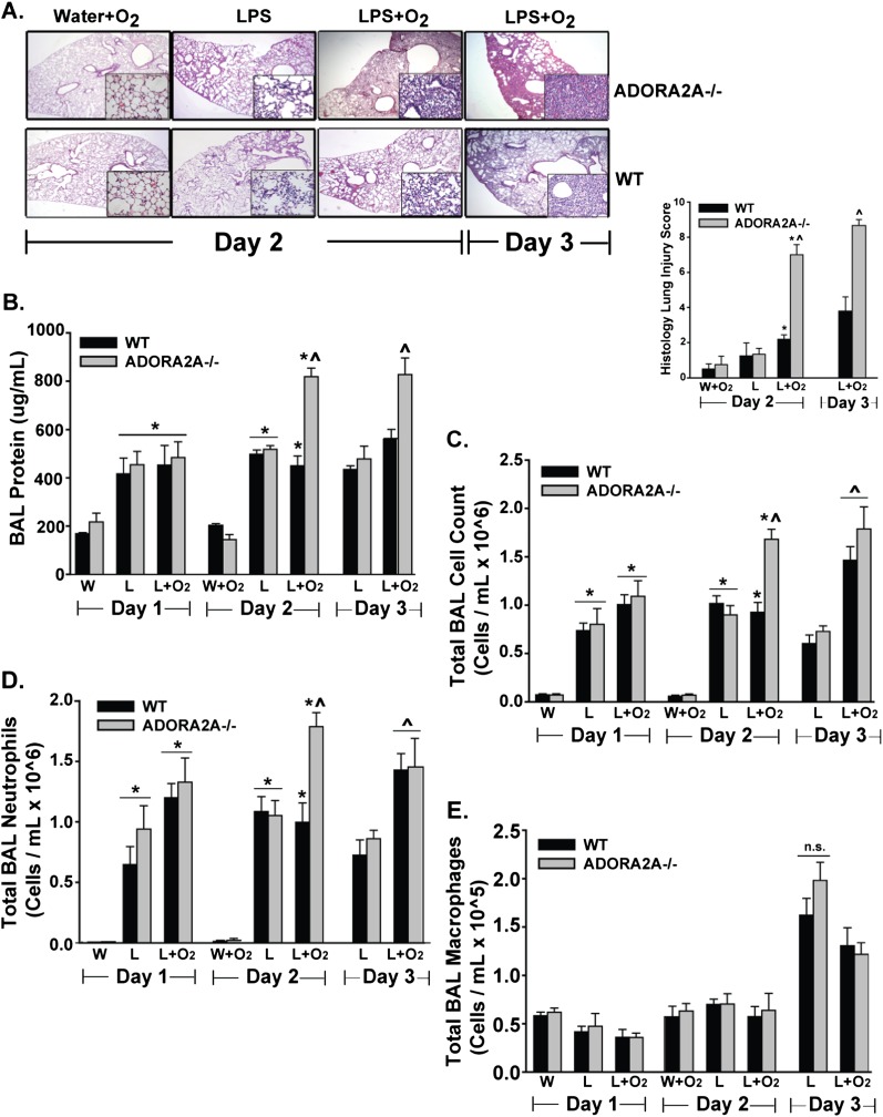
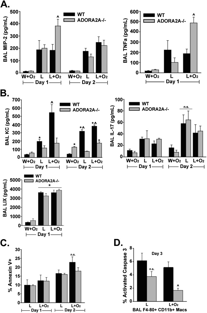
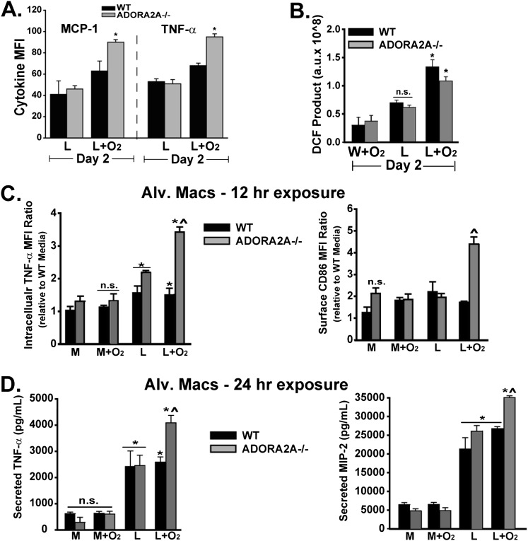
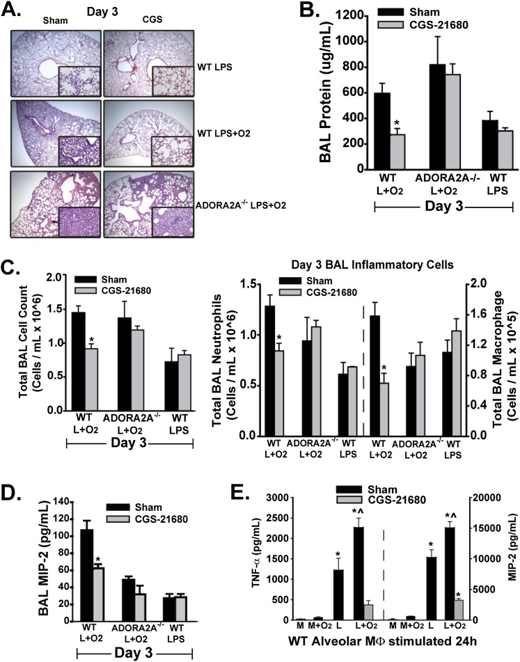
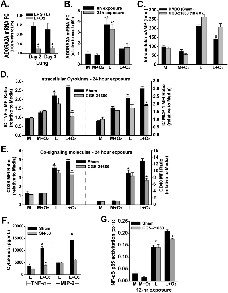
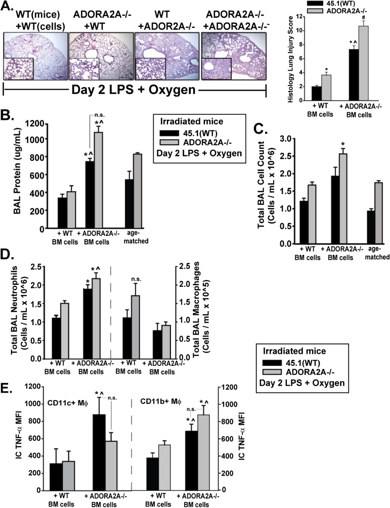
Acute respiratory distress syndrome (ARDS) causes significant morbidity and mortality. Exacerbating factors increasing the risk of ARDS remain unknown. Supplemental oxygen is often necessary in both mild and severe lung disease. The potential effects of supplemental oxygen may include augmentation of lung inflammation by inhibiting anti-inflammatory pathways in alveolar macrophages. We sought to determine oxygen-derived effects on the anti-inflammatory A2A adenosinergic (ADORA2A) receptor in macrophages, and the role of the ADORA2A receptor in lung injury. Wild-type (WT) and ADORA2A(-/-) mice received intratracheal lipopolysaccharide (IT LPS), followed 12 hours later by continuous exposure to 21% oxygen (control mice) or 60% oxygen for 1 to 3 days. We measured the phenotypic endpoints of lung injury and the alveolar macrophage inflammatory state. We tested an ADORA2A-specific agonist, CGS-21680 hydrochloride, in LPS plus oxygen-exposed WT and ADORA2A(-/-) mice. We determined the specific effects of myeloid ADORA2A, using chimera experiments. Compared with WT mice, ADORA2A(-/-) mice exposed to IT LPS and 60% oxygen demonstrated significantly more histologic lung injury, alveolar neutrophils, and protein. Macrophages from ADORA2A(-/-) mice exposed to LPS plus oxygen expressed higher concentrations of proinflammatory cytokines and cosignaling molecules. CGS-21680 prevented the oxygen-induced augmentation of lung injury after LPS only in WT mice. Chimera experiments demonstrated that the transfer of WT but not ADORA2A(-/-) bone marrow cells into irradiated ADORA2A(-/-) mice reduced lung injury after LPS plus oxygen, demonstrating myeloid ADORA2A protection. ADORA2A is protective against lung injury after LPS and oxygen. Oxygen after LPS increases macrophage activation to augment lung injury by inhibiting the ADORA2A pathway.


Figures

<i>Figure 1.</i>
Figure 1.
Anti-inflammatory A2A adenosinergic receptor–deficient (ADORA2A−/−) mice exposed to LPS plus oxygen demonstrated a significant increase in lung injury compared with ADORA2A−/− mice exposed to LPS alone, and compared with wild-type (WT) mice exposed to LPS plus oxygen. Statistics were performed by comparing all groups at the same time point. (A) Histology. Representative low-power (×4) and high-power (inset, ×40) hematoxylin and eosin–stained lung sections on Days 2 and 3 reveal increased alveolar consolidation and interstitial infiltration in ADORA2A−/− mice exposed to LPS plus oxygen. Quantitative assessment using a lung-injury scoring system demonstrated a significant increase in lung injury in ADORA2A−/− mice exposed to LPS plus oxygen, beginning on Day 2 and persisting until Day 3. *On Day 2, significant compared with the WT water (W) + O2 group. ^On Day 2, significant compared with all other groups. ^On Day 3, significant compared with the WT LPS (L) + O2 group. (B) Bronchoalveolar lavage (BAL) protein was notable for an early, pronounced increase in ADORA2A−/− mice exposed to LPS plus oxygen. *On Day 1, significant compared with the WT W and ADORA2A−/− W groups. *On Day 2, significant compared with the WT W + O2 and ADORA2A−/− W + O2 groups. ^On Day 2, significant compared with the WT LPS, WT L + O2, and ADORA2A−/− LPS groups. ^On Day 3, significant compared with the WT LPS, WT L + O2, and ADORA2A−/− LPS groups. (C) The BAL total cell count was increased in ADORA2A−/− mice exposed to LPS plus oxygen, compared with all other exposure groups on Day 2. *On Day 1, significant compared with the WT W and ADORA2A−/− W groups. *On Day 2, significant compared with the WT W + O2 and ADORA2A−/− W + O2 groups. ^On Day 2, significant compared with the WT LPS, WT L + O2, and ADORA2A−/− LPS groups. ^On Day 3, significant compared with the WT LPS and ADORA2A−/− LPS groups. (D) BAL Neutrophils were also increased in ADORA2A−/− mice exposed to LPS plus oxygen on Day 2. *On Day 1, significant compared with the WT W and ADORA2A−/− W groups. *On Day 2, significant compared with the WT W + O2 and ADORA2A−/− W + O2 groups. ^On Day 2, significant compared with the WT LPS, WT L + O2, and ADORA2A−/− LPS groups. ^On Day 3, significant compared with the WT LPS and ADORA2A−/− LPS groups. (E) BAL macrophages did not increase in ADORA2A−/− mice exposed to LPS plus oxygen, compared with all other groups at each time point (n = 4 in the W and W + O2 groups; n = 6–8 in the LPS and L + O2 groups). n.s., no significance.
<i>Figure 2.</i>
Figure 2.
Macrophages from ADORA2A−/− mice secrete more neutrophil-recruiting chemokines and are less apoptotic after exposure to LPS plus oxygen, compared with WT mice. Statistics were determined by comparing all groups at the same time point. (A) BAL concentrations of macrophage inflammatory protein–2 (MIP-2) and TNF-α were increased in ADORA2A−/− mice exposed to LPS plus oxygen on Day 1. ^Significant compared with the WT LPS, WT L + O2, and ADORA2A−/− LPS groups. (B) Other chemokines, including keratinocyte-derived chemokine (KC), IL-17, and LPS-induced chemokine (LIX), were not increased in ADORA2A−/− mice exposed to LPS plus oxygen, compared with WT mice. ^Significant compared with all other groups on BAL KC Day 1. *Significant compared with the WT W + O2 group on BAL KC Day 1. *Significant compared with the WT W + O2 group on BAL KC Day 2. ^Significant compared with all other groups on BAL KC Day 2. For BAL IL-17, no difference was evident between the assessed groups. *For BAL LIX, significant compared with the WT and ADORA2A−/− W + O2 groups. (C) Amounts of annexin V, a marker of BAL neutrophil apoptosis, were no different between any of the assessed groups on either Day 2 or Day 3. (D) Amounts of activated caspase-3, a marker of BAL macrophage (F4-80+ CD11b+) apoptosis, were significantly lower in ADORA2A−/− mice exposed to LPS plus oxygen. ^Significant difference compared with the WT LPS, WT L + O2, and ADORA2A−/− LPS groups, and with no difference between the WT LPS and ADORA2A−/− LPS groups (n = 4 in the W and W + O2 groups, and n = 6–8 in the LPS and L + O2 groups). Macs, macrophages.
<i>Figure 3.</i>
Figure 3.
ADORA2A-deficient macrophages exposed to LPS plus oxygen demonstrate increased cytokine production as well as cosignaling molecule expression, compared with WT macrophages. Statistics were performed by comparing all groups at the same time point. (A) MCP-1 and TNF-α were increased in macrophages (F4-80+ CD11b+) from ADORA2A−/− mice exposed to LPS plus oxygen. *Significant compared with the WT LPS, ADORA2A−/− LPS, and WT L + O2 groups. MFI, mean fluorescence intensity. (B) As a marker of reactive oxygen species (ROS) production, the 2′,7′-dichlorofluorescein (DCF) measured in alveolar macrophages was higher in both WT and ADORA2A−/− mice exposed to LPS and oxygen. *Significant compared with all other groups, with nonsignificant differences between the WT L + O2 and ADORA2A−/− L + O2 groups. (C) Using alveolar macrophages (Alv Macs.) isolated from unchallenged WT or ADORA2A−/− mice and exposed in culture to LPS, 60% oxygen, or the two in combination, we observed the most significant increase in TNF-α and CD86 in macrophages from ADORA2A−/− mice exposed to LPS and oxygen after 12 hours. *For intracellular (IC) TNF-α, significant compared with WT media (M). ^For IC TNF-α, significant compared with the WT LPS, WT L + O2, and ADORA2A−/− LPS groups, with no difference between the ADORA2A−/− LPS, WT LPS, or WT L + O2 groups. ^For CD86, significant compared with the WT LPS, WT L + O2, and ADORA2A−/− LPS groups. (D) When assessed after 24 hours of exposure, macrophages from ADORA2A−/− mice exposed to LPS plus oxygen secreted more TNF-α and MIP-2. *Significant compared with WT media. ^Significant compared with the WT LPS, WT L + O2, and ADORA2A−/− LPS groups (n = 5–6 in each group).
<i>Figure 4.</i>
Figure 4.
CGS-21680 hydrochloride, an ADORA2A-specific agonist, abrogates lung injury in WT mice induced by LPS plus oxygen. To do so, CGS-21680 prevents the oxygen-mediated suppression of the ADORA2A pathway. Statistics were determined by comparing groups with and without CGS-21680 treatment. (A) Representative low-power (×4) and high-power (inset, ×40) hematoxylin and eosin–stained lung sections on Day 3 after intratracheal (IT) LPS demonstrate a CGS-21680–mediated reduction in histologic injury in WT mice exposed to LPS plus oxygen. (B) BAL protein is significantly reduced with the addition of CGS-21680 to WT mice exposed to LPS plus oxygen *Significant compared with the WT L + O2 group. No measureable CGS-21680–mediated difference was evident in WT mice exposed to LPS alone, or in ADORA2A−/− mice exposed to LPS plus oxygen. (C) Adding CGS-21680 to LPS plus oxygen–exposed WT mice significantly reduced total BAL cells as well as BAL neutrophils and macrophages. *Significant compared with the WT L + O2 group. (D) CGS-21680 blunted the LPS plus oxygen–mediated increase in BAL MIP-2. *Significant compared with the WT L + O2 group. (E) We isolated naive, WT alveolar macrophages (Mφ) and exposed them in culture to LPS, oxygen, or both in combination for 24 hours. Statistics were determined by comparing all groups at the same time point. Adding CGS-21680 to the LPS plus oxygen–exposed macrophages significantly blunted the 24-hour TNF-α and MIP-2 secretion. *Significant compared with the media (M) and M + O2 groups. ^Significant compared with the LPS and L + O2 + CGS-21680 groups, with no significant difference between the LPS and L + O2 + CGS-21680 groups (n = 5–6 in each group).
<i>Figure 5.</i>
Figure 5.
Oxygen blunts the induction by LPS of ADORA2A and ADORA2A pathway components in macrophages to augment the proinflammatory profile. This effect is negated by the ADORA2A agonist, CGS-21680. Statistics were determined by comparing all groups at the same time point. (A) Adding oxygen to LPS reduced lung ADORA2A mRNA concentrations on Days 2 and 3. *Significant compared with LPS-treated mice at the same time point. FC, fold change. (B) LPS induced an early and sustained increase in ADORA2A mRNA in murine alveolar macrophage cell line (MH-S) cells, an effect negated by the addition of oxygen. *Significant compared with the media and M + O2 groups. ^Significant compared with the L + O2 8-hour and L + O2 24-hour groups. (C) Oxygen significantly reduced the LPS-mediated increase in intracellular cyclic adenosine monophosphate (IC cAMP), an effect negated by treatment with CGS-21680. *Significant compared with the LPS + O2 + CGS-21680 group. n.s., between the LPS and LPS + CGS-21680 groups. (D) Adding CGS-21680 to LPS plus oxygen–treated cells significantly reduced IC TNF-α and IC MCP-1 production to concentrations comparable to those with LPS treatment alone. *Significant compared with the L + O2 group. (E) Adding CGS-21680 to LPS plus oxygen–treated cells significantly reduced the CD86 and CD40 mean fluorescence intensity (MFI) compared with LPS treatment alone. *Significant compared with the L + O2 group. (F) Adding the NF-κB inhibitor, cell permeable synthetic peptide (SN-50) (50 μg/ml), at the time of oxygen exposure (3 hours after LPS) most significantly reduced the macrophage secretion of TNF-α and MIP-2 in LPS plus oxygen–exposed cells at 24 hours. *Significant compared with the LPS + SN-50 group. ^Significant compared with all other groups for each cytokine. A small reduction in TNF-α secretion by LPS-treated cells was evident when SN-50 was added, but with no significant change in MIP-2. (G) LPS plus oxygen exposure significantly increased NF-κB activity in macrophages compared with LPS alone. Adding CGS-21680 to the LPS plus oxygen–treated group led to a 50% reduction in the increase in NF-κB activity observed with the addition of oxygen to LPS versus LPS alone. *Significant compared with the L + O2 group, with n.s. between the L + O2 + CGS-21680 and LPS or LPS + CGS-21680 groups (n = 5–6 in each group).
<i>Figure 6.</i>
Figure 6.
Chimera experiments demonstrate protective effects of myeloid and nonmyeloid ADORA2A in the LPS plus oxygen lung-injury model. Statistics were determined by comparing all groups at the same time point. All mice were exposed to LPS plus oxygen for 2 days. (A) Representative low-power (×4) and high-power (inset, ×40) hematoxylin and eosin–stained lung sections on Day 2 after delivery of IT LPS plus oxygen to defined chimera groups demonstrate increased alveolar consolidation in mice with ADORA2A−/− myeloid cells. Quantitative assessment, based on a lung-injury scoring system, demonstrated maximal histologic injury in ADORA2A−/− mice that received ADORA2A−/− myeloid cells, with the next most severe injury in the WT mice that received ADORA2A−/− myeloid cells, compared with other groups. *Significant increase in lung injury compared with the WT mice + WT cell group. ^Significant increase compared with the ADORA2A−/− mice + WT cell group. #Significant increase compared with all three groups. (B) Concentrations of BAL protein were significantly higher in WT and ADORA2A−/− mice that received ADORA2A−/− myeloid cells. *Significant compared with the WT mice + WT cell group. ^Significant compared with the ADORA2A−/− mice + WT cell group. (C) BAL total cell count was significantly increased in ADORA2A−/− mice that received ADORA2A−/− cells. *Significant only compared with the WT mice + WT cell and the ADORA2A−/− mice + WT cell groups. (D) BAL neutrophils were significantly increased in both groups that received ADORA2A−/− myeloid cells when assessed on Day 2. *Significant compared with the WT mice + WT cell group. ^Significant compared with the ADORA2A−/− mice + WT cell group. (E) Among ex vivo alveolar macrophages (F4-80+ CD11c+ and F4-80+ CD11b+), we observed a significant increase in IC TNF-α production when assessed by flow cytometry. *Significant compared with the WT mice + WT cell group. ^Significant compared with the ADORA2A−/− mice + WT cell group (n = 4 in each group). BM, bone marrow.

Similar articles

Cited by

References

    1. Matthay M, Zimmerman G. Acute lung injury and the acute respiratory distress syndrome: four decades of inquiry into pathogenesis and rational management. Am J Respir Cell Mol Biol. 2005;33:319–327. - PMC - PubMed
    1. Gajic O, Dabbagh O, Park PK, Adesanya A, Chang SY, Hou P, Anderson H, Hoth JJ, Mikkelsen ME, Gentile NT, et al. Early identification of patients at risk of acute lung injury: evaluation of lung injury prediction score in a multicenter cohort study. Am J Respir Crit Care Med. 2011;183:462–470. - PMC - PubMed
    1. Matute-Bello G, Frevert CW, Martin TR. Animal models of acute lung injury. Am J Physiol Lung Cell Mol Physiol. 2008;295:L379–L399. - PMC - PubMed
    1. Bertolini G, Lewandowski K, Bion J, Romand JA, Villar J, Thorsteinsson A, Damas P, Armaganidis A, Lemaire F, Minelli C, et al. Epidemiology and outcome of acute lung injury in European intensive care units. Intensive Care Med. 2004;30:51–61. - PubMed
    1. Pepe PE, Potkin RT, Reus DH, Hudson LD, Carrico CJ. Clinical predictors of the adult respiratory distress syndrome. Am J Surg. 1982;144:124–130. - PubMed

Publication types

MeSH terms