Review
. 2013 Apr;34(2):209-38.
doi: 10.1210/er.2012-1015. Epub 2013 Jan 24.

Recent insights into the cell biology of thyroid angiofollicular units

Affiliations
Review

Recent insights into the cell biology of thyroid angiofollicular units

Ides M Colin et al. Endocr Rev. 2013 Apr.

Abstract

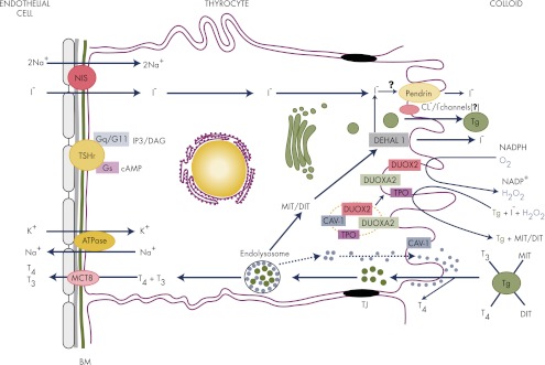
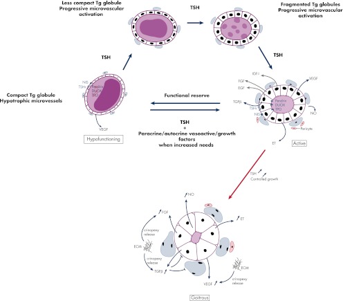
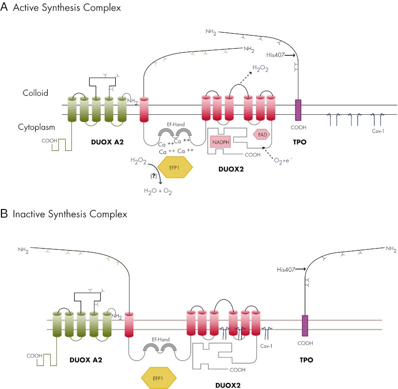
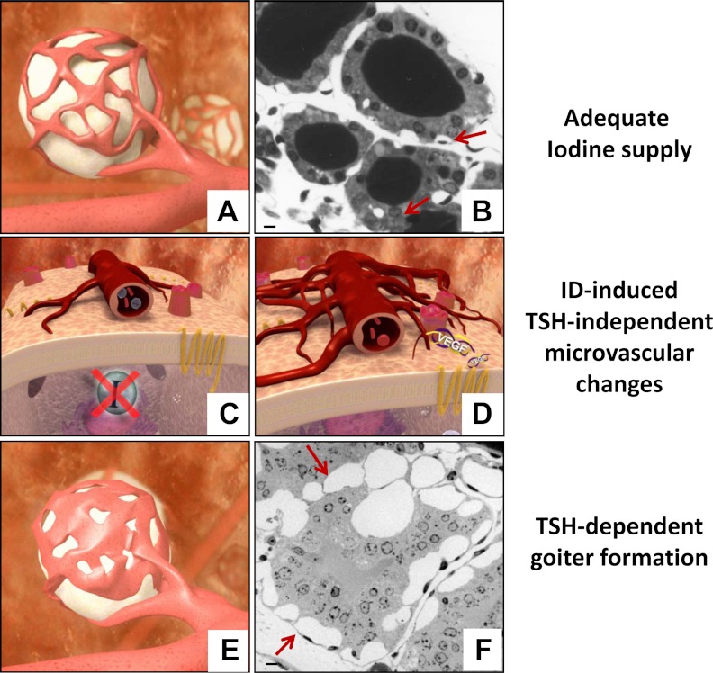
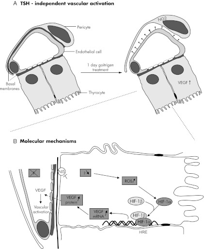
In thyrocytes, cell polarity is of crucial importance for proper thyroid function. Many intrinsic mechanisms of self-regulation control how the key players involved in thyroid hormone (TH) biosynthesis interact in apical microvilli, so that hazardous biochemical processes may occur without detriment to the cell. In some pathological conditions, this enzymatic complex is disrupted, with some components abnormally activated into the cytoplasm, which can lead to further morphological and functional breakdown. When iodine intake is altered, autoregulatory mechanisms outside the thyrocytes are activated. They involve adjacent capillaries that, together with thyrocytes, form the angiofollicular units (AFUs) that can be considered as the functional and morphological units of the thyroid. In response to iodine shortage, a rapid expansion of the microvasculature occurs, which, in addition to nutrients and oxygen, optimizes iodide supply. These changes are triggered by angiogenic signals released from thyrocytes via a reactive oxygen species/hypoxia-inducible factor/vascular endothelial growth factor pathway. When intra- and extrathyrocyte autoregulation fails, other forms of adaptation arise, such as euthyroid goiters. From onset, goiters are morphologically and functionally heterogeneous due to the polyclonal nature of the cells, with nodules distributed around areas of quiescent AFUs containing globules of compact thyroglobulin (Tg) and surrounded by a hypotrophic microvasculature. Upon TSH stimulation, quiescent AFUs are activated with Tg globules undergoing fragmentation into soluble Tg, proteins involved in TH biosynthesis being expressed and the local microvascular network extending. Over time and depending on physiological needs, AFUs may undergo repetitive phases of high, moderate, or low cell and tissue activity, which may ultimately culminate in multinodular goiters.


Figures

Figure 1.
Figure 1.
The intrathyroidal journey of iodide. Iodide (I) entering the thyroid gland through fenestrated microvessels must sequentially cross the two basal laminae of endothelial and epithelial cells to be transported into the thyrocyte by NIS, which uses the energy provided by a Na+-K+-ATPase. Once inside the cell, I rapidly goes to the apical surface (according to a process that still remains elusive) where it is transported across the apical membrane by pendrin, even though other apical transporters have been proposed to be part of this process. I is readily oxidized by TPO in the presence of H2O2 and incorporated into tyrosine residues of Tg to form MIT and DIT, which are coupled, again by TPO, to form T3 and T4. H2O2 is produced by DUOX2 in coordination with DUOXA2. TH biosynthesis occurs at the interface with the colloid in a harmless environment for the cell. Iodinated Tg is stored in the colloid until use (see Fig. 2). T4 can be partly released extracellularly by cathepsins K and L. In the human, the uptake of Tg by thyrocytes occurs by micropinocytosis, which can be either nonspecific (fluid phase) or receptor mediated. After endocytosis, colloid droplets containing partly digested Tg are transported to the endolysosomal compartment for complete hydrolysis by lysosomal enzymes including cathepsins. THs are released into the circulation via basal TH transporters (including MCT8) and transported to their target tissues via binding transport proteins. Unused MIT and DIT are dehalogenated by DEHAL1, and most of the I is recycled into TH synthesis. The binding of TSH to its receptor activates both Gs and Gq proteins. Green dots represent colloid droplets with partially degraded Tg, and blue dots represent cathepsins. BM, Basal membranes; DAG, diacylglycerol; Gq/G11, guanine nucleotide-binding protein αq and α11 subunits; Gs, guanine nucleotide-binding protein α-subunit; IP3, inositol triphosphate; TJ, tight junctions; TSHr, TSH receptor.
Figure 2.
Figure 2.
Proposed mechanism of hypofunctioning follicle reactivation and processing of Tg globules upon TSH stimulation. In the normal human thyroid, active follicles coexist with many hypofunctioning follicles that are surrounded by hypotrophic capillaries and that contain compact Tg globules. These globules correspond to condensed Tg in covalently cross-linked form, not immediately available for TH synthesis, but eventually usable in case of increased hormonal needs. When this occurs, hypofunctioning follicles serve as a functional reserve that can be reactivated upon TSH stimulation. TPO, DUOX, and pendrin expression then reemerges along with progressive dilution of Tg globules into soluble Tg, under the action of secreted cathepsins. This occurs along with the expansion of adjacent microvessels, which, besides TSH, depends also upon many locally secreted growth and vasoactive factors. Persistent TSH stimulation induces thyrocyte hypertrophy and proliferation resulting in goitrous follicles with no more colloid Tg reserves. The microvascular reaction keeps evolving with the proliferation of endothelial cells, the fusion of adjacent microvessels, and the formation of larger microvessels. At this stage, in addition to being secreted by thyrocytes, VEGF, as well as FGF at least, could also function as crinopexins that are locally synthesized molecules sequestered in pericellular structures (extracellular matrix, cell surface, and proteoglycans) under a latent form (305). According to this theory, early alterations in local blood flow and in microvessel shape may alter the extracellular matrix, thereby provoking the release and the activation of crinopexins that could, in turn, stimulate endothelial cell proliferation. This would amplify local changes in the microvasculature initially originated from thyrocytes. Of note, VEGF expression is observed even in thyrocytes of hypofunctioning follicles. ECM, Extracellular matrix; ET, endothelins; TSHr, TSH receptor.
Figure 3.
Figure 3.
The multiprotein complex involved in TH biosynthesis. A, Upon TSH stimulation, DUOX2 and TPO migrate into the plasma membrane where the complex is activated. TPO and DUOX2 are viewed as a producer-consumer unit that allows TPO to use H2O2 instantly to oxidize iodide (I) into iodine, to iodinate tyrosyl residues of Tg, and to couple iodotyrosyl residues to form T3 and T4, while protecting the cell against H2O2 possible oxidative damage. DUOX2 is represented here as a seven-transmembrane-domain glycoprotein with an extracellular NH2-terminal peroxidase-like domain that appears essential for interaction with TPO, a long COOH-terminal region that contains the catalytic NADPH oxidase core along with the flavin adenine dinucleotide (FAD) and NADPH binding cavities, and two EF-hands in the first intracellular loop that are essential calcium binding sites. TPO is represented with a short intracellular COOH terminus and a long extracellular NH2 terminus (90% of its amino acids) that exhibits a catalase-like activity in certain circumstances. His407 is involved in the covalent binding of the heme prosthetic group that is essential for enzyme activity. The manner in which TPO and DUOX2 interact makes the apical multiprotein complex perfectly suited to detoxify ROS and avoid H2O2 spillages. The local H2O2 concentration is kept under control by the TPO catalase-like action (which protects DUOX2, as long as both proteins are closely associated). In addition, the complex contains the thioredoxin-related protein EFP1, which interacts with the two EF-hands of DUOX2 and detoxifies H2O2 that is not readily used in the biosynthesis process. EFP1 may also be involved in the maturation process of DUOX2. For this biochemical unit to be activated, DUOX2 must be fully glycosylated (Y, N-glycosylation sites) following successive maturation steps in the ER and the Golgi apparatus. DUOX2 must also be associated with the maturation factor DUOXA2, which is represented here as a five-transmembrane-domain protein with an extracellular loop between the second and the third transmembrane domains along with three glycosylation sites (Y). I and H2O2 regulate the activity of the enzymatic complex, depending on their local relative concentration. When I is present in adequate amounts, H2O2 is the limiting factor of the reaction and mediates the association between TPO and DUOX2. In thyroids with low intracellular I content, I instead exerts a stimulatory effect on H2O2 production. When I is present in excess, H2O2 synthesis is blocked. Because I-induced effects are inhibited by methimazole or propylthiouracil, this indicates that I acts through oxidized species. B, In resting conditions, the complex TPO-DUOX2-DUOXA2 is kept inactive underneath the apical membrane. Cav-1 is required to keep DUOX2 quiescent, because it may interact with domains in the cytoplasmic region of the molecule that are putative sites for Cav-1 scaffolding domains (following a hypothesis that still needs to be proved). According to the most plausible explanation, Cav-1 would come off the enzyme complex after its incorporation into the apical membrane. The absence of Cav-1 is responsible for mislocalization and premature activation of the complex in the cytoplasm, creating potentially devastating consequences due to increased OS and/or lack of efficient antioxidant mechanisms naturally activated when I organification and coupling reactions normally occur in apical microvilli.
Figure 4.
Figure 4.
The concept of AFUs in the thyroid gland. The thyroid gland is composed of many AFUs. They are individualized entities with their own genotypic and phenotypic assets under the local control of a host of autocrine and paracrine factors, as well as exerted by TSH. A and B, Each unit is composed of thyrocytes gathered in a follicle that is surrounded by capillaries made of endothelial cells and pericytes independent from their neighbors. In unstimulated conditions (i.e. in conditions of adequate iodine supply), the vascular bed covers about 20–50% of the follicle surface (this AFU corresponds to the so-called active follicle in Fig. 2). C and D, As soon as the intracellular iodide (I) content drops, the thyrocytes react immediately by secreting VEGF, which triggers an early and modest TSH-independent microvascular reaction. If the increased local clearance of I associated with this compensatory mechanism is not enough to preserve TH synthesis, TSH plasma levels start rising. Upon TSH stimulation, a more robust but always tightly controlled microvascular reaction occurs. E and F, By the end of the process, the area occupied by the expanded vascular bed may cover up to 80% of the follicle surface made of hyperplastic and hypertrophic thyrocytes. Scale bar, 10 μm. Arrows indicate microvessels.
Figure 5.
Figure 5.
Proposed mechanism of early ID-induced TSH-independent microvascular reaction in thyroid AFUs. A, TSH-independent microvascular activation. In unstimulated AFUs (left), capillaries close to thyrocytes are fenestrated and surrounded by pericytes. Endothelial cells and pericytes are separated only by fused basal membranes. If iodide (I) supply drops, independently from any stimulation by TSH (right), VEGF expression increases in thyrocytes, which induces a microvascular activation, implying, as initial step, the detachment of chondroitin sulfate proteoglycan 4 (NG2)-expressing pericytes from endothelial cells. B, Molecular mechanisms. When Isupply is altered or when its transport is blocked, the thyrocyte intracellular content drops rapidly, thereby increasing ROS, which leads to the stabilization of HIF-1α. The exact nature of ROS and where they are produced still deserve further investigation. HIF-1α heterodimerizes with HIF-1β, which is constitutively present in the cell. The HIF-1α/HIF-1β heterodimer, after binding to the hypoxia response element (HRE) site of the promoter region of the VEGF gene, turns on its transcription. As a result, VEGF mRNA and protein expression increases. VEGF is then secreted and activates adjacent endothelial cells and pericytes, thereby leading to microvascular activation, increased blood flow, and proliferation of endothelial cells and, in turn, to increased local clearance of I. When this and other protective measures become insufficient to safeguard TH synthesis, TSH enters into the action, in coordination with many other locally generated growth and vascular regulatory factors.

References

    1. Boelaert K, Franklyn JA. 2005. Thyroid hormone in health and disease. J Endocrinol 187:1–15 - PubMed
    1. Zimmermann MB. 2011. The role of iodine in human growth and development. Semin Cell Dev Biol 22:645–652 - PubMed
    1. Zimmermann MB. 2009. Iodine deficiency. Endocr Rev 30:376–408 - PubMed
    1. Courtois B. 1813. Découverte d'une substance nouvelle dans le Vareck. Ann Chim (Paris) 88:304–310
    1. Taurog A. 2004. Iodine. In: Martini L, ed. Encyclopedia of endocrine diseases. New York: Academic Press; 82–88

MeSH terms