

The trithorax protein partner menin acts in tandem with EZH2 to suppress C/EBPα and differentiation in MLL-AF9 leukemia
- PMID: 23349306
- PMCID: PMC3669449
- DOI: 10.3324/haematol.2012.074195
The trithorax protein partner menin acts in tandem with EZH2 to suppress C/EBPα and differentiation in MLL-AF9 leukemia
Abstract
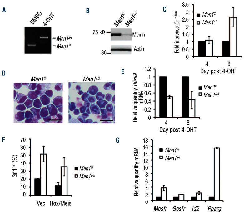
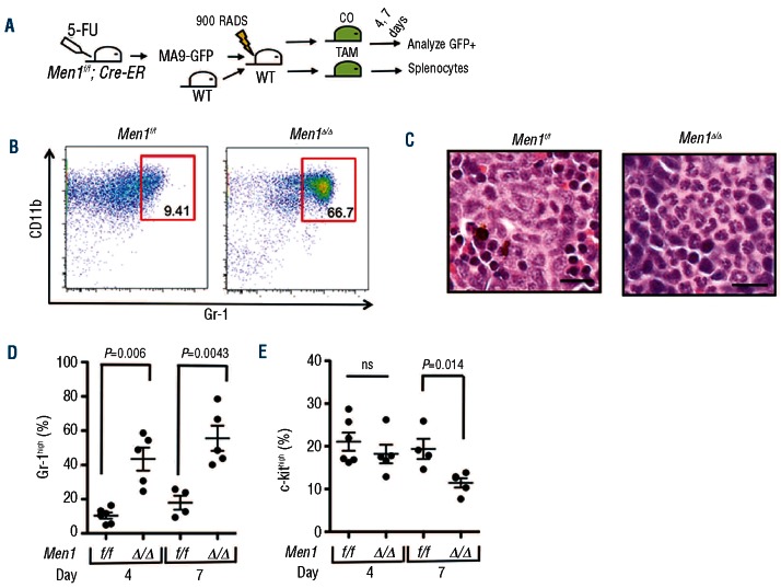
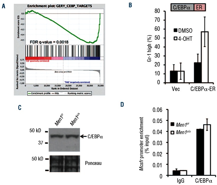
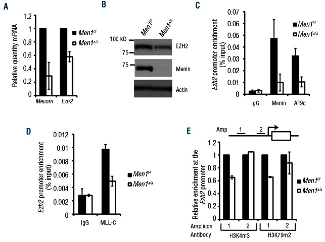
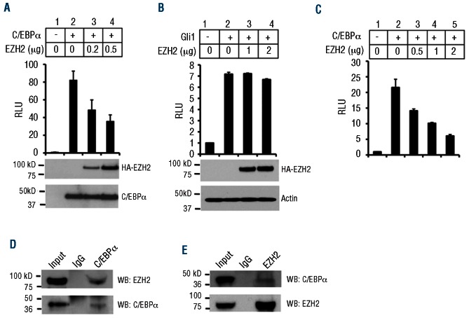
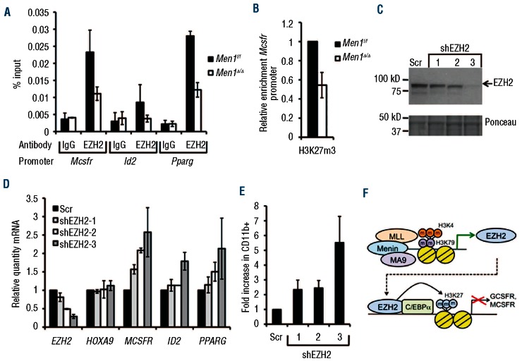
Trithorax and polycomb group proteins antagonistically regulate the transcription of many genes, and cancer can result from the disruption of this regulation. Deregulation of trithorax function occurs through chromosomal translocations involving the trithorax gene MLL, leading to the expression of MLL fusion proteins and acute leukemia. It is poorly understood how MLL fusion proteins block differentiation, a hallmark of leukemogenesis. We analyzed the effect of acute depletion of menin, a close partner of MLL that is critical for MLL and MLL-AF9 recruitment to target genes, on MLL-AF9 leukemia cell differentiation using an in vivo model. We performed cDNA microarray analysis of menin-regulated genes from primary leukemia cells to determine menin-regulated pathways involved in suppressing MLL-AF9 leukemia cell differentiation. We found that menin binds the promoter of the polycomb gene Ezh2, and promotes its expression. EZH2 interacts with the differentiation-promoting transcription factor C/EBPα and represses C/EBPα target genes. Menin depletion reduces MLL binding to the Ezh2 locus, EZH2 expression, and EZH2 binding and repressive H3K27 methylation at C/EBPα target genes, thereby inducing the expression of pro-differentiation C/EBPα targets. In conclusion, our results show that in contrast to its classical role antagonizing trithorax function, the polycomb group protein EZH2 collaborates with trithorax-associated menin to block MLL-AF9 leukemia cell differentiation, uncovering a novel mechanism for suppression of C/EBPα and leukemia cell differentiation, through menin-mediated upregulation of EZH2.
Figures
Comment in
-
Trithorax and polycomb cooperation in MLL fusion acute leukemia.Haematologica. 2013 Jun;98(6):825-7. doi: 10.3324/haematol.2013.084301. Haematologica. 2013. PMID: 23729722 Free PMC article. No abstract available.
Similar articles
-
Trithorax and polycomb cooperation in MLL fusion acute leukemia.Haematologica. 2013 Jun;98(6):825-7. doi: 10.3324/haematol.2013.084301. Haematologica. 2013. PMID: 23729722 Free PMC article. No abstract available.
-
MLL-AF9-induced leukemogenesis requires coexpression of the wild-type Mll allele.Cancer Cell. 2010 Feb 17;17(2):148-59. doi: 10.1016/j.ccr.2009.12.034. Cancer Cell. 2010. PMID: 20159607 Free PMC article.
-
Regulation of HOXA9 activity by predominant expression of DACH1 against C/EBPα and GATA-1 in myeloid leukemia with MLL-AF9.Biochem Biophys Res Commun. 2012 Sep 28;426(3):299-305. doi: 10.1016/j.bbrc.2012.08.048. Epub 2012 Aug 17. Biochem Biophys Res Commun. 2012. PMID: 22902925
-
Menin as a hub controlling mixed lineage leukemia.Bioessays. 2012 Sep;34(9):771-80. doi: 10.1002/bies.201200007. Epub 2012 Jul 24. Bioessays. 2012. PMID: 22829075 Free PMC article. Review.
-
Disordered epigenetic regulation in MLL-related leukemia.Int J Hematol. 2012 Oct;96(4):428-37. doi: 10.1007/s12185-012-1180-0. Epub 2012 Sep 29. Int J Hematol. 2012. PMID: 23054645 Review.
Cited by
-
Trithorax and polycomb cooperation in MLL fusion acute leukemia.Haematologica. 2013 Jun;98(6):825-7. doi: 10.3324/haematol.2013.084301. Haematologica. 2013. PMID: 23729722 Free PMC article. No abstract available.
-
Menin: a scaffold protein that controls gene expression and cell signaling.Trends Biochem Sci. 2013 Aug;38(8):394-402. doi: 10.1016/j.tibs.2013.05.005. Epub 2013 Jul 10. Trends Biochem Sci. 2013. PMID: 23850066 Free PMC article. Review.
-
A Review of the Scaffold Protein Menin and its Role in Hepatobiliary Pathology.Gene Expr. 2017 Jul 7;17(3):251-263. doi: 10.3727/105221617X695744. Epub 2017 Apr 28. Gene Expr. 2017. PMID: 28485270 Free PMC article. Review.
-
Menin in Cancer.Genes (Basel). 2024 Sep 21;15(9):1231. doi: 10.3390/genes15091231. Genes (Basel). 2024. PMID: 39336822 Free PMC article. Review.
-
Disruption of the menin-MLL interaction triggers menin protein degradation via ubiquitin-proteasome pathway.Am J Cancer Res. 2019 Aug 1;9(8):1682-1694. eCollection 2019. Am J Cancer Res. 2019. PMID: 31497350 Free PMC article.
References
-
- Schuettengruber B, Martinez AM, Iovino N, Cavalli G. Trithorax group proteins: switching genes on and keeping them active. Nat Rev Mol Cell Biol. 2011;12(12):799–814 - PubMed
-
- Pineault N, Helgason CD, Lawrence HJ, Humphries RK. Differential expression of Hox, Meis1, and Pbx1 genes in primitive cells throughout murine hematopoietic ontogeny. Exp Hematol. 2002;30(1):49–57 - PubMed
-
- Milne TA, Briggs SD, Brock HW, Martin ME, Gibbs D, Allis CD, et al. MLL targets SET domain methyltransferase activity to Hox gene promoters. Mol Cell. 2002;10(5):1107–17 - PubMed
Publication types
MeSH terms
Substances
Grants and funding
LinkOut - more resources
Full Text Sources
Other Literature Sources
Medical
