. 2013 Jan;9(1):e1003125.
doi: 10.1371/journal.ppat.1003125. Epub 2013 Jan 22.

The hepatitis B virus ribonuclease H is sensitive to inhibitors of the human immunodeficiency virus ribonuclease H and integrase enzymes

Affiliations

The hepatitis B virus ribonuclease H is sensitive to inhibitors of the human immunodeficiency virus ribonuclease H and integrase enzymes

John E Tavis et al. PLoS Pathog. 2013 Jan.

Abstract

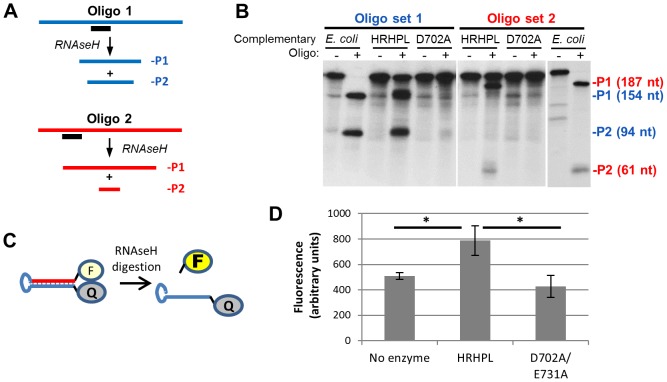
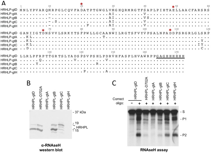
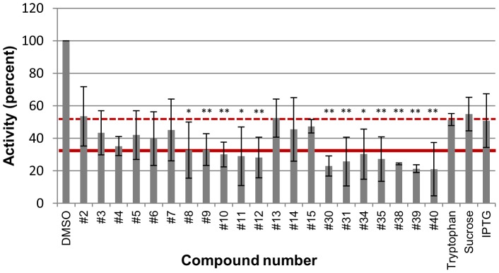
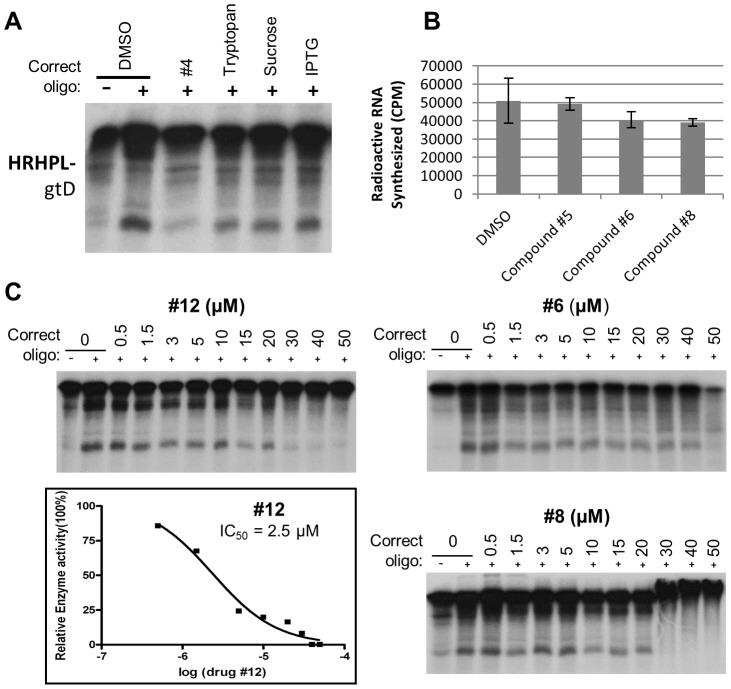
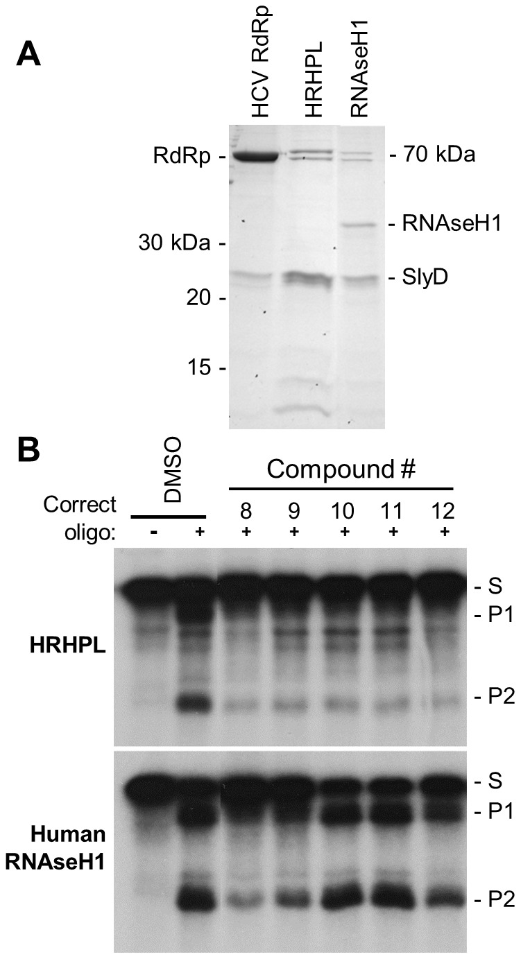
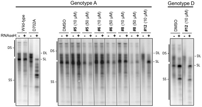
Nucleos(t)ide analog therapy blocks DNA synthesis by the hepatitis B virus (HBV) reverse transcriptase and can control the infection, but treatment is life-long and has high costs and unpredictable long-term side effects. The profound suppression of HBV by the nucleos(t)ide analogs and their ability to cure some patients indicates that they can push HBV to the brink of extinction. Consequently, more patients could be cured by suppressing HBV replication further using a new drug in combination with the nucleos(t)ide analogs. The HBV ribonuclease H (RNAseH) is a logical drug target because it is the second of only two viral enzymes that are essential for viral replication, but it has not been exploited, primarily because it is very difficult to produce active enzyme. To address this difficulty, we expressed HBV genotype D and H RNAseHs in E. coli and enriched the enzymes by nickel-affinity chromatography. HBV RNAseH activity in the enriched lysates was characterized in preparation for drug screening. Twenty-one candidate HBV RNAseH inhibitors were identified using chemical structure-activity analyses based on inhibitors of the HIV RNAseH and integrase. Twelve anti-RNAseH and anti-integrase compounds inhibited the HBV RNAseH at 10 µM, the best compounds had low micromolar IC(50) values against the RNAseH, and one compound inhibited HBV replication in tissue culture at 10 µM. Recombinant HBV genotype D RNAseH was more sensitive to inhibition than genotype H. This study demonstrates that recombinant HBV RNAseH suitable for low-throughput antiviral drug screening has been produced. The high percentage of compounds developed against the HIV RNAseH and integrase that were active against the HBV RNAseH indicates that the extensive drug design efforts against these HIV enzymes can guide anti-HBV RNAseH drug discovery. Finally, differential inhibition of HBV genotype D and H RNAseHs indicates that viral genetic variability will be a factor during drug development.


Conflict of interest statement

I have read the journal's policy and have the following conflicts: JET will be an inventor on a pending patent application covering this RNAseH assay and the compounds identified with it. SGS and MAP will be inventors on patent application(s) covering compounds #12-15. This does not alter our adherence to all PLoS Pathogens policies on sharing data and materials.

Figures

Figure 1
Figure 1. The HBV replication cycle.
HBV replicates by reverse transcription in the cytoplasm of infected hepatocytes. After completion of reverse transcription, intracellular capsids can either be transported into the nucleus to maintain the cccDNA pool (Recycling), or they can be enveloped and secreted from the cells as mature virions (Secretion). Inhibiting RNAseH activity blocks plus-strand DNA synthesis during reverse transcription; this would prevent both recycling and secretion of virions. The hepatocyte is represented as a rectangle, the nucleus as an oval, HBV capsids as a hexagon, and the viral lipid envelop as a circle surrounding the extracellular capsids. HBV proteins are green or black, RNAs are red, and DNAs are blue.
Figure 2
Figure 2. Alignments between the HBV RNAseH and the HIV-1 RNAseH and integrase.
Manually optimized alignments between HBV RNAseH and A. the HIV-1 RNAseH, or B. the HIV-1 integrase. The HBV genotype D sequence is from Genbank entry V01460 and the HIV-1 sequences are from strain HXB2; Genbank K03455.1. Identical residues are shaded in black and similar residues are shaded in gray. * indicates the conserved nucleotidly transferase superfamily active site carboxylates (D-E-D-D for the RNAseH enzymes and D-D-E for the integrase). The numbering for each sequence is indicated at top. Residue 1 for the HBV RNAseH domain is amino acid 684 in the full-length polymerase protein (strain adw2) and residue 1 for the HIV-1 RNAseH domain is amino acid 441 of the full-length reverse transcriptase (strain HXB2).
Figure 3
Figure 3. Identification of the DEDD motif in the HBV RNAseH active site.
Wild-type and mutant HBV genotype A genomic expression vectors were transfected into cells, intracellular capsids were isolated five days later, and viral nucleic acids were purified from the capsids. The nucleic acids were divided into two aliquots; one aliquot was treated with DNAse-free E. coli RNAseH to destroy RNA:DNA heteroduplexes and the other was mock treated. The nucleic acids were resolved by agarose electrophoresis and HBV DNAs were detected by Southern analysis. The signature of an RNAseH-deficient genome is production of RNA:DNA heteroduplexes in which the DNA migrates as double-stranded species without treatment with exogenous RNAseH treatment but as singe-stranded species following degradation of the RNA. The positions of the duplex linear (DL) and full-length single-stranded linear (SL) HBV DNA markers are shown. DS indicates the spectrum of double-stranded nucleic acids produced by reverse transcription, and SS indicates the spectrum of single-stranded nucleic acids.
Figure 4
Figure 4. Recombinant HBV RNAseH proteins.
A. Structure of the recombinant RNAseHs. The HBV polymerase with its major domains labeled is at top. The recombinant RNAseH derivatives are shown below with the C-terminal hexahistidine tag in brown. TP, terminal protein domain; RT, reverse transcriptase domain; *, mutations D702A or E731A to RNAseH active site residues. B. Proteins in the enriched lysates. The left panel is a Coomassie-blue stained SDS-PAGE gel of enriched RNAseH extracts as employed in the RNAseH assays. The right panel is a western blot of the extracts employing monoclonal antibody 9F9 which recognizes an epitope near the C-terminus of the HBV polymerase.
Figure 5
Figure 5. Recombinant HBV RNAseH is enzymatically active.
A. Oligonucleotide-directed RNAseH assay. Uniformly 32P-labeled RNA (blue or red) is annealed to a complementary DNA oligonucleotide (black). RNAseH activity cleaves the RNA in the heteroduplex formed where the oligonucleotide anneals to the RNA and yields two products (P1 and P2). B. Recombinant HBV RNAseH is active. An oligonucleotide-directed RNAseH assay was conducted with E. coli RNAseH, wild-type HBV RNAseH (HRHPL), or RNAseH-deficient HRHPL (D702A). A complementary oligonucleotide (+) or non-complementary oligonucleotide (−) was mixed with labeled DRF+ RNA and the reactions were incubated to allow RNAseH activity. The products were resolved by SDS-PAGE and the RNAs were detected by autoradiography. Oligonucleotide set 1 was D2507− and D2526+ and oligonucleotide set #2 was D2543M-Sal and D2453+. The positions of the cleavage products (P1 and P2) are indicated in blue for reactions containing oligonucleotide D2507− and in red for reactions containing oligonucleotide D2543M-Sal. C. FRET-based RNAseH assay. A self-complementary chimeric RNA:DNA synthetic oligonucleotide (RHF1) forms a stem-loop in which the stem is an RNA:DNA heteroduplex. The stem brings the fluorescein (F) and quencher (Q) at the 5′ and 3′ ends of the oligonucleotide into close proximity. Cleavage of the RNA releases the fluorescein and increases its fluorescence. D. Detection of HBV RNAseH activity employing the fluorescent assay. The substrate in panel C was employed in an RNAseH assay employing buffer alone, wild-type HBV RNAseH (HRHPL), or RNAseH-deficient HRHPL (D702A/E731A). *, P<0.05.
Figure 6
Figure 6. Recombinant RNAseHs from HBV genotypes A, B, C, D, and H.
A. Sequence alignment for genotype A, B, C, D, and H versions of the HBV RNAseH expression construct HRHPL. The additional methionine at residue 10 of the genotype D sequence is a product of the cloning strategy; this insertion has no impact on the RNAseH activity because the first 9 amino acids of HRHPL can be deleted without altering the biochemical profile of the enzyme. * indicates the DEDD active site residues, and the hexahistidine tag at the C-terminus is underlined. Residue 1 for the HBV RNAseH domain is amino acid 684 in the full-length polymerase protein (strain adw2). B. Western analysis of RNAseH proteins in the enriched lysates probed with the anti-HBV RNAseH monoclonal antibody 9F9. C. RNAseH activity of RNAseH from HBV genotypes A, B, C, D, and H detected by the oligonucleotide-directed RNA cleavage assay. HRHPL-D702A (genotype D) is a negative control. gt, genotype.
Figure 7
Figure 7. Inhibition of the HBV RNAseH by candidate compounds selected for their similarity to antagonists of the HIV RNAseH and integrase.
Candidate inhibitors (compounds #2-40) and irrelevant compounds (tryptophan, sucrose, and IPTG) were included at 10 µM in a standard oligonucleotide-directed RNAseH assay employing wild-type genotype D HBV RNAseH (HRHPL). DMSO, vehicle control. Error bars are ± one standard deviation from three to seven replicates. The dashed red line indicates the mean residual activity in the irrelevant control reactions (52%) and the solid red line is two standard deviations of the irrelevant control assays below their mean (33%). Compounds that inhibited the RNAseH to 33% or below were considered to be positive (“+” in Table 2). *, P<0.05 by T-test against the pooled data for the irrelevant controls; **, P<0.01.
Figure 8
Figure 8. Specificity of anti-HBV RNAseH compounds.
A. Inhibition of HBV genotype D RNAseH by irrelevant compounds at 10 µM in the oligonucleotide-directed RNAseH assay. Compound #4 was employed as an example HBV RNAseH inhibitor. B. Anti-HBV RNAseH inhibitors do not significantly inhibit the HCV RNA polymerase. The ability of compounds #5, 6 and 8 to inhibit production of poly-G by the HCV RNA-directed RNA polymerase was measured in a primed homopolymeric RNA synthesis assay . The compounds were employed at 10 µM. DMSO, vehicle control. C. Dose-responsiveness of HBV RNAseH inhibition. The effects of compounds #6, 8, and 12 on the RNAseH activity of HRHPL (genotype D) were measured at concentrations ranging from 0.5 to 50 µM. The dose-response profile is plotted for compound #12.
Figure 9
Figure 9. Activity of HBV RNAseH inhibitors against human RNAseH1.
A. Proteins in the enriched recombinant human RNAseH1 lysates employed in the RNAseH reactions were detected by Coomassie-blue staining following SDS-PAGE. B. An oligonucleotide-directed RNAseH assay was conducted with wild-type HBV RNAseH (genotype D) and recombinant human RNAseH1 under identical reaction conditions. The inhibitory compounds were employed at 10 µM. The upper and lower panels are from the same experiment and the data were collected on a single sheet of film, so the reactions can be directly compared. DMSO, vehicle control. S, the DRF+ substrate; P1 and P2, RNAseH cleavage products.
Figure 10
Figure 10. Inhibition of HBV replication in culture by RNAseH inhibitors.
Genotype A or D HBV genomic expression vectors were transfected into cells, intracellular capsids were isolated four days later, and viral nucleic acids were purified from the capsids. The nucleic acids were divided into two aliquots; one aliquot was treated with DNAse-free E. coli RNAseH to destroy RNA:DNA heteroduplexes and the other was mock treated. The nucleic acids were resolved by agarose electrophoresis and HBV DNAs were detected by Southern analysis. Inhibition of RNAseH activity leads to accumulation of RNA:DNA heteroduplexes in which the DNA migrates as double-stranded species in the mock-treated sample but as faster-migrating singe-stranded species following RNAseH treatment. The left panel is a control in which wild-type and RNAseH-deficient D702A HBV genomes were compared. The right two panels employed wild-type HBV in the presence of the test compounds. Compounds were employed at 10 or 50 µM as indicated. DMSO, vehicle control. The positions of the duplex linear (DL) and full-length single-stranded linear (SL) HBV DNA markers are shown. DS indicates the spectrum of double-stranded nucleic acids produced by reverse transcription, and SS indicates the spectrum of single-stranded nucleic acids.

Similar articles

Cited by

References

    1. Seeger C, Zoulim F, Mason WS (2007) Hepadnaviruses. In: Knipe DM, Howley P, Griffin DE, Lamb RA, Martin MA et al..., editors. Fields Virology. Philadelphia: Lippincott Williams & Wilkins. pp. 2977–3029.
    1. Shepard CW, Simard EP, Finelli L, Fiore AE, Bell BP (2006) Hepatitis B virus infection: epidemiology and vaccination. Epidemiol Rev 28: 112–125. - PubMed
    1. Ganem D, Prince AM (2004) Hepatitis B virus infection–natural history and clinical consequences. N Engl J Med 350: 1118–1129. - PubMed
    1. Lavanchy D (2004) Hepatitis B virus epidemiology, disease burden, treatment, and current and emerging prevention and control measures. J Viral Hepat 11: 97–107. - PubMed
    1. Tavis JE, Badtke MP (2009) Hepadnaviral Genomic Replication. In: Cameron CE, Götte M, Raney KD, editors. Viral Genome Replication. New York: Springer Science+Business Media, LLC. pp. 129–143.

Publication types

MeSH terms

Substances