Clinical Trial
. 2013;8(1):e53629.
doi: 10.1371/journal.pone.0053629. Epub 2013 Jan 17.

Analysis of V2 antibody responses induced in vaccinees in the ALVAC/AIDSVAX HIV-1 vaccine efficacy trial

Affiliations
Clinical Trial

Analysis of V2 antibody responses induced in vaccinees in the ALVAC/AIDSVAX HIV-1 vaccine efficacy trial

Susan Zolla-Pazner et al. PLoS One. 2013.

Abstract

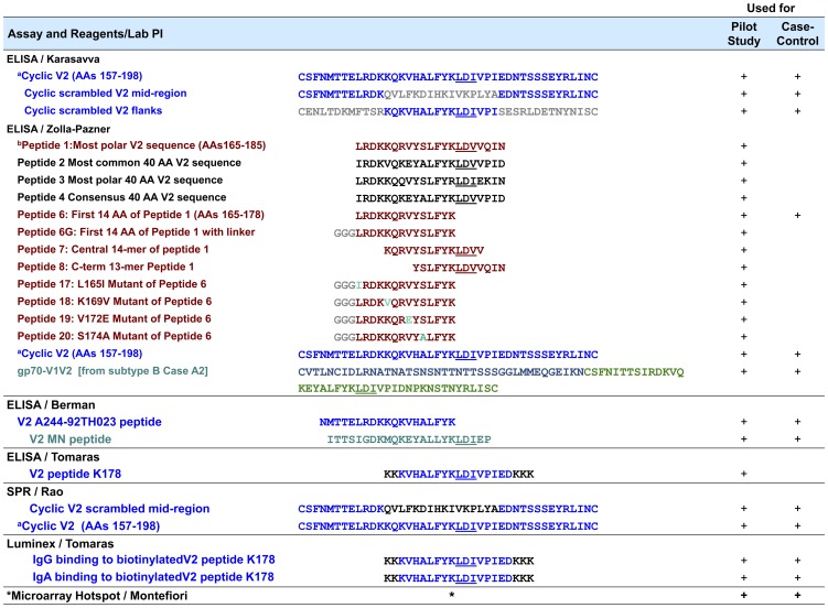
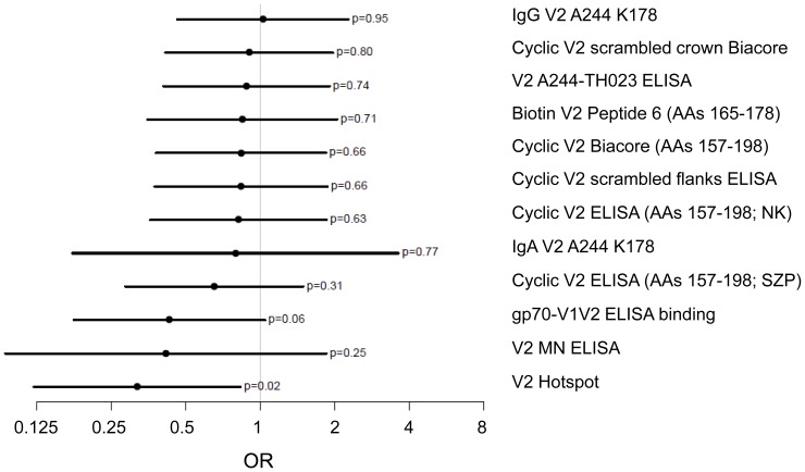
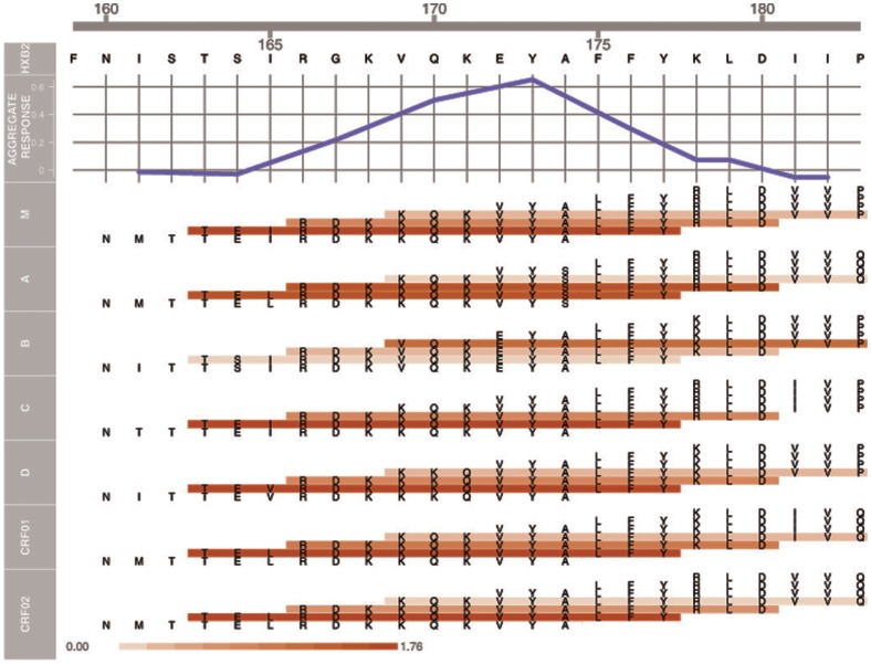
The RV144 clinical trial of a prime/boost immunizing regimen using recombinant canary pox (ALVAC-HIV) and two gp120 proteins (AIDSVAX B and E) was previously shown to have a 31.2% efficacy rate. Plasma specimens from vaccine and placebo recipients were used in an extensive set of assays to identify correlates of HIV-1 infection risk. Of six primary variables that were studied, only one displayed a significant inverse correlation with risk of infection: the antibody (Ab) response to a fusion protein containing the V1 and V2 regions of gp120 (gp70-V1V2). This finding prompted a thorough examination of the results generated with the complete panel of 13 assays measuring various V2 Abs in the stored plasma used in the initial pilot studies and those used in the subsequent case-control study. The studies revealed that the ALVAC-HIV/AIDSVAX vaccine induced V2-specific Abs that cross-react with multiple HIV-1 subgroups and recognize both conformational and linear epitopes. The conformational epitope was present on gp70-V1V2, while the predominant linear V2 epitope mapped to residues 165-178, immediately N-terminal to the putative α4β7 binding motif in the mid-loop region of V2. Odds ratios (ORs) were calculated to compare the risk of infection with data from 12 V2 assays, and in 11 of these, the ORs were ≤1, reaching statistical significance for two of the variables: Ab responses to gp70-V1V2 and to overlapping V2 linear peptides. It remains to be determined whether anti-V2 Ab responses were directly responsible for the reduced infection rate in RV144 and whether anti-V2 Abs will prove to be important with other candidate HIV vaccines that show efficacy, however, the results support continued dissection of Ab responses to the V2 region which may illuminate mechanisms of protection from HIV-1 infection and may facilitate the development of an effective HIV-1 vaccine.


Conflict of interest statement

Competing Interests: The authors have declared that no competing interests exist.

Figures

Figure 1
Figure 1. V2-related Reagents and Assays Used in the Pilot and Case-Control Analyses.
The superscripts denote the following: a “Cyclic V2 (AAs 157–198)” was used in assays in three labs as shown. Throughout this table, blue V2 sequences are identical to the subtype E 92TH023 used in the prime; gray represent scrambled sequences or linkers; brown represents the sequence the central AAs in an extremely polar V2 in subtype A strain QB585.2102M.Ev1v5.C (http://www.hiv.lanl.gov) with individual mutations shown in cyan; black represents sequences chosen for particular properties, as described; green represents the V1V2 from subtype B Case A2 and the central 23-mer of V2 from subtype B strain MN. b All peptides were biotinylated at the N-terminus with the exception of peptide K178 and peptide V2 A244-92TH023 which were biotinylated at the C-terminus. * Multiple V2 peptides from various strains (see text).
Figure 2
Figure 2. ELISA reactivity of human anti-V2 mAbs 697-D and 2158 with AIDSVAX subtype E (A244, green square) and AIDSVAX subtype B (MN, O) gp120 immunogens.
The pink dashed line represents twice background levels.
Figure 3
Figure 3. ELISA reactivity with gp70-V1V2 of plasma specimens used in the pilot studies of the RV144 clinical vaccine trial (Set C).
The results from one of three experiments are shown. The open and filled blue diamonds depict negative responses at weeks 0 and 26, respectively. The open and closed red circles depict positive responses at weeks 0 and 26, respectively. Each vertical line connects a single patient's specimen drawn at Week 0 and Week 26. The specimens are ordered by the difference in reactivity between the Week 0 and Week 26 specimens, with the biggest increasers on the right. Plasma were tested at a final dilution of 1∶100, and a positive response was defined as being >0.283, the cut-off OD value which was defined as the mean +3 standard deviations based on values derived from vaccinees at week 0 (the pre-immunization time point).
Figure 4
Figure 4. Spearman rank correlations between the V2 assays run in the case-control study.
Figure 5
Figure 5. Boxplots showing ELISA reactivity of V2 peptides with plasma (diluted 1∶100) from 80 vaccinees' specimens from the pilot study (Set C).
The distributions of the reactivities are shown where the left edge of each box equals the 25th percentile; the vertical line in each box is the 50th percentile, and the right edge of each box equals the 75th percentile. The boxplots were prepared using Prism, with “whiskers” showing the minimum and maximum responses. Panel A: Four 21-mer N-terminal biotinylated peptides (Peptides 1–4) were selected on the basis of a bioinformatics analysis of V2 sequences from the LANL HIV Database (see text). Panel B: A second peptide panel designed upon inspection of the AAs in Peptides 1–4 in Panel A revealed AAs that distinguish Peptides 1 and 3 from 2 and 4; these appear at positions 165, 169, 172, and 174. To maximally enhance the availability of the epitopes on the peptides used in the fine mapping of the V2 Abs, a spacer of three glycines was inserted between the biotin tag at the N-terminus of the peptide and the V2 sequences.
Figure 6
Figure 6. Ribbon diagram of the backbone fold of the V1V2 domain (purple and yellow ribbons) bound to the CDR H3 region of mAb PG9 (transparent stippled spheres are the atoms of the PG9 CDR H3, blue = nitrogen, red = oxygen, white = carbon).
The ribbon backbone of AAs 165–176, which are identical to Peptide 6 (in Figure 5 ), is colored yellow. The strands are labeled A–D according to the convention established recently .
Figure 7
Figure 7. Estimated odds ratios (OR) and 95% confidence intervals for each of the V2 assays.
Data are derived from the categorical analyses shown in Table 1 . Estimated ORs compare subgroups defined by High vs. Low responses except for comparisons for analyses of IgA V2 A244 K178 and V2 MN which compare Positive vs. Negative responses. For evaluation of biotinylated V2 Peptide 6, comparison is between High and Negative responses.
Figure 8
Figure 8. Hotspot analysis of the microarray data of V2 Abs in the plasma from vaccinees in the case-control study.
The upper panel shows an aggregate response across all sub-types averaged across vaccinees as a function of the V2 sequence in subtype B strain HxB2. An individual aggregate response is computed using a sliding window mean statistic of 9 AAs, i.e., peptides with HxB2 positions of 9 contiguous AAs averaged together. In the lower portion of the figure, the actual sequence of each of the overlapping 15-mers spanning V2 positions 160–183 is shown. The seven sets of peptides represent the consensus V2 regions of HIV-1 Group M and of subtypes A, B, C, D, CRF01_AE and CRF02_AG. Peptides are colored according to their average response across all vaccinees, with a scale of 0 (white) to 1.76 (red). All responses are calculated using normalized intensities and by subtracting the intensities of baseline pre-bleeds.

Similar articles

Cited by

References

    1. Rerks-Ngarm S, Pitisuttithum P, Nitayaphan S, Kaewkungwal J, Chiu J, et al. (2009) Vaccination with ALVAC and AIDSVAX to Prevent HIV-1 Infection in Thailand. N Engl J Med 361: 2209–2220. - PubMed
    1. Haynes BF, Gilbert PB, McElrath MJ, Zolla-Pazner S, Tomaras GD, et al. (2012) Immune Correlates Analysis of the ALVAC-AIDSVAX HIV-1 Vaccine Efficacy Trial. N Engl J Med 366: 1275–1286. - PMC - PubMed
    1. Zolla-Pazner S, Cardozo T (2010) Structure-Function Relationships of HIV-1 Envelope Sequence-Variable Regions Provide a Paradigm for Vaccine Design. Nat Rev Immunol 10: 527–535. - PMC - PubMed
    1. Gorny MK, Xu J-Y, Karwowska S, Buchbinder A, Zolla-Pazner S (1993) Repertoire of neutralizing human monoclonal antibodies specific for the V3 domain of HIV-1 gp120. J Immunol 150: 635–643. - PubMed
    1. Israel ZR, Gorny MK, Palmer C, McKeating JA, Zolla-Pazner S (1997) Prevalence of a V2 epitope in clade B primary isolates and its recognition by sera from HIV-1 infected individuals. Aids 11: 128–130. - PubMed

Publication types