

Choroidal thickness and biometric markers for the screening of lacquer cracks in patients with high myopia
- PMID: 23349728
- PMCID: PMC3551908
- DOI: 10.1371/journal.pone.0053660
Choroidal thickness and biometric markers for the screening of lacquer cracks in patients with high myopia
Abstract
Objectives: Validation of choroidal thickness and other biometrics measured by spectral domain optical coherence tomography (SD-OCT) in predicting lacquer cracks formation in highly myopic eyes.
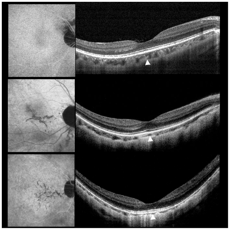
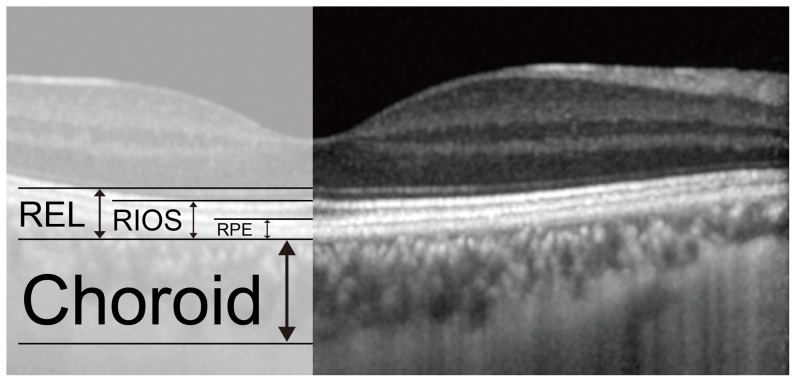
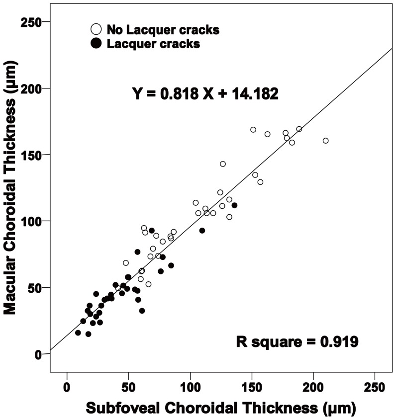
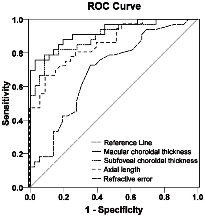
Methods: Patients with a refractive error worse than -8 diopters and moderate myopic maculopathy were recruited into two groups based on the presence or absence of lacquer cracks (36 eyes without and 33 eyes with lacquer cracks). Choroidal thickness, refractive error, and axial length were measured and subjected to receiver operating characteristic curve analysis to identify the optimal cutoff values at predicting lacquer crack formation. The width of the retinal pigment epithelium (RPE), RPE to the inner segment/outer segment line, RPE to the external limiting membrane were also measured and compared to the subfoveal choroidal thickness to assess their relationships as potential markers of lacquer crack formation.
Results: Lacquer crack is associated with decreased choroidal thickness, lower best-corrected visual acuity, longer axial length and higher refractive errors. Choroidal thickness has the strongest association with lacquer crack formation versus axial length and refractive error. In eyes with lacquer cracks, stellate lacquer cracks are associated with thinner choroidal thickness compared to eyes with linear lacquer cracks. Subfoveal choroidal thickness less than the width of the retinal pigment epithelium to the inner segment/outer segment line is also associated with lacquer crack formation (sensitivity 78.8%, specificity 88.3%, and accuracy 81.2%).
Conclusions: This study suggests that choroidal thickness and other SD-OCT measurements could be employed clinically to predict the development and severity of lacquer cracks in patients with high myopia.
Conflict of interest statement
Figures
References
-
- Xu L, Wang Y, Li Y, Cui T, Li J, et al. (2006) Causes of blindness and visual impairment in urban and rural areas in Beijing: the Beijing Eye Study. Ophthalmology 113: 1134 e1–11. - PubMed
-
- Iwase A, Araie M, Tomidokoro A, Yamamoto T, Shimizu H, et al. (2006) Prevalence and causes of low vision and blindness in a Japanese adult population: the Tajimi Study. Ophthalmology 113: 1354–1362. - PubMed
-
- Hsu WM, Cheng CY, Liu JH, Tsai SY, Chou P (2004) Prevalence and causes of visual impairment in an elderly Chinese population in Taiwan: the Shihpai Eye Study. Ophthalmology 111: 62–69. - PubMed
-
- Liu JH, Cheng CY, Chen SJ, Lee FL (2001) Visual impairment in a Taiwanese population: prevalence, causes, and socioeconomic factors. Ophthalmic Epidemiol 8: 339–350. - PubMed
Publication types
MeSH terms
Substances
Grants and funding
LinkOut - more resources
Full Text Sources
Other Literature Sources
