

Staphylococcus aureus and Pseudomonas aeruginosa express and secrete human surfactant proteins
- PMID: 23349731
- PMCID: PMC3551896
- DOI: 10.1371/journal.pone.0053705
Staphylococcus aureus and Pseudomonas aeruginosa express and secrete human surfactant proteins
Abstract
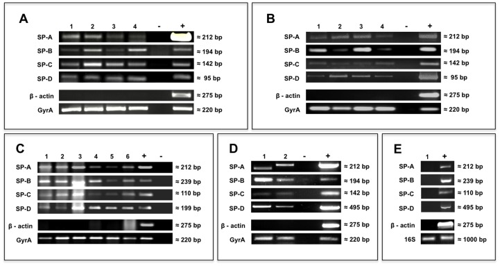
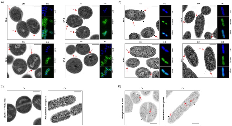
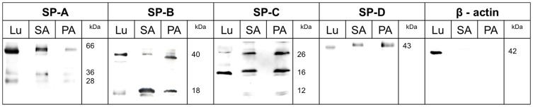
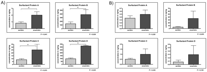
Surfactant proteins (SP), originally known from human lung surfactant, are essential to proper respiratory function in that they lower the surface tension of the alveoli. They are also important components of the innate immune system. The functional significance of these proteins is currently reflected by a very large and growing number of publications. The objective goal of this study was to elucidate whether Staphylococcus aureus and Pseudomonas aeruginosa is able to express surfactant proteins. 10 different strains of S. aureus and P. aeruginosa were analyzed by means of RT-PCR, Western blot analysis, ELISA, immunofluorescence microscopy and immunoelectron microscopy. The unexpected and surprising finding revealed in this study is that different strains of S. aureus and P. aeruginosa express and secrete proteins that react with currently commercially available antibodies to known human surfactant proteins. Our results strongly suggest that the bacteria are either able to express 'human-like' surfactant proteins on their own or that commercially available primers and antibodies to human surfactant proteins detect identical bacterial proteins and genes. The results may reflect the existence of a new group of bacterial surfactant proteins and DNA currently lacking in the relevant sequence and structure databases. At any rate, our knowledge of human surfactant proteins obtained from immunological and molecular biological studies may have been falsified by the presence of bacterial proteins and DNA and therefore requires critical reassessment.
Conflict of interest statement
Figures
Similar articles
-
Pattern differentiation in co-culture biofilms formed by Staphylococcus aureus and Pseudomonas aeruginosa.FEMS Immunol Med Microbiol. 2011 Aug;62(3):339-47. doi: 10.1111/j.1574-695X.2011.00820.x. Epub 2011 Jun 14. FEMS Immunol Med Microbiol. 2011. PMID: 21595754
-
Heme protects Pseudomonas aeruginosa and Staphylococcus aureus from calprotectin-induced iron starvation.J Biol Chem. 2021 Jan-Jun;296:100160. doi: 10.1074/jbc.RA120.015975. Epub 2020 Dec 9. J Biol Chem. 2021. PMID: 33273016 Free PMC article.
-
Accumulation of 99mTc-ciprofloxacin in Staphylococcus aureus and Pseudomonas aeruginosa.Antimicrob Agents Chemother. 2008 Jul;52(7):2691-2. doi: 10.1128/AAC.00217-08. Epub 2008 May 12. Antimicrob Agents Chemother. 2008. PMID: 18474577 Free PMC article.
-
Pseudomonas aeruginosa and Staphylococcus aureus communication in biofilm infections: insights through network and database construction.Crit Rev Microbiol. 2019 Sep-Nov;45(5-6):712-728. doi: 10.1080/1040841X.2019.1700209. Epub 2019 Dec 13. Crit Rev Microbiol. 2019. PMID: 31835971 Review.
-
Chronic wound infections: the role of Pseudomonas aeruginosa and Staphylococcus aureus.Expert Rev Anti Infect Ther. 2015 May;13(5):605-13. doi: 10.1586/14787210.2015.1023291. Epub 2015 Mar 8. Expert Rev Anti Infect Ther. 2015. PMID: 25746414 Review.
Cited by
-
Immunolocalization of Surfactant Proteins SP-A, SP-B, SP-C, and SP-D in Infantile Labial Glands and Mucosa.J Histochem Cytochem. 2018 Jul;66(7):531-538. doi: 10.1369/0022155418766063. Epub 2018 Mar 30. J Histochem Cytochem. 2018. PMID: 29601229 Free PMC article.
-
Structure, genetics and function of the pulmonary associated surfactant proteins A and D: The extra-pulmonary role of these C type lectins.Ann Anat. 2017 May;211:184-201. doi: 10.1016/j.aanat.2017.03.002. Epub 2017 Mar 27. Ann Anat. 2017. PMID: 28351530 Free PMC article. Review.
-
The detection of surfactant proteins A, B, C and D in the human brain and their regulation in cerebral infarction, autoimmune conditions and infections of the CNS.PLoS One. 2013 Sep 30;8(9):e74412. doi: 10.1371/journal.pone.0074412. eCollection 2013. PLoS One. 2013. PMID: 24098648 Free PMC article.
-
The novel surfactant protein SP-H enhances the phagocytosis efficiency of macrophage-like cell lines U937 and MH-S.BMC Res Notes. 2014 Nov 26;7:851. doi: 10.1186/1756-0500-7-851. BMC Res Notes. 2014. PMID: 25427765 Free PMC article.
-
Collagen and hyaluronan at wound sites influence early polymicrobial biofilm adhesive events.BMC Microbiol. 2014 Jul 16;14:191. doi: 10.1186/1471-2180-14-191. BMC Microbiol. 2014. PMID: 25026865 Free PMC article.
References
-
- Von Neegaard K (1929) Neue Auffassungen über einen Grundbegriff der Atemmechanik. ZGesamte Exp Med 66: 373–394.
-
- King RJ, Klass DJ, Gikas EG, Clements JA (1973) Isolation of apoproteins from canine surface active material. The American journal of physiology 224: 788–795. - PubMed
-
- Van Iwaarden F, Welmers B, Verhoef J, Haagsman HP, van Golde LM (1990) Pulmonary surfactant protein A enhances the host-defense mechanism of rat alveolar macrophages. Am J Respir Cell Mol Biol 2: 91–98. - PubMed
-
- Yu SH, Possmayer F (1990) Role of bovine pulmonary surfactant-associated proteins in the surface-active property of phospholipid mixtures. Biochimica et biophysica acta 1046: 233–241. - PubMed
MeSH terms
Substances
LinkOut - more resources
Full Text Sources
Other Literature Sources
Molecular Biology Databases
