. 2013;8(1):e53781.
doi: 10.1371/journal.pone.0053781. Epub 2013 Jan 21.

Cloning, annotation and developmental expression of the chicken intestinal MUC2 gene

Affiliations

Cloning, annotation and developmental expression of the chicken intestinal MUC2 gene

Zhengyu Jiang et al. PLoS One. 2013.

Abstract

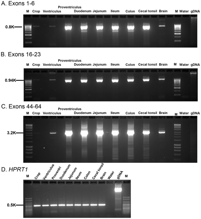
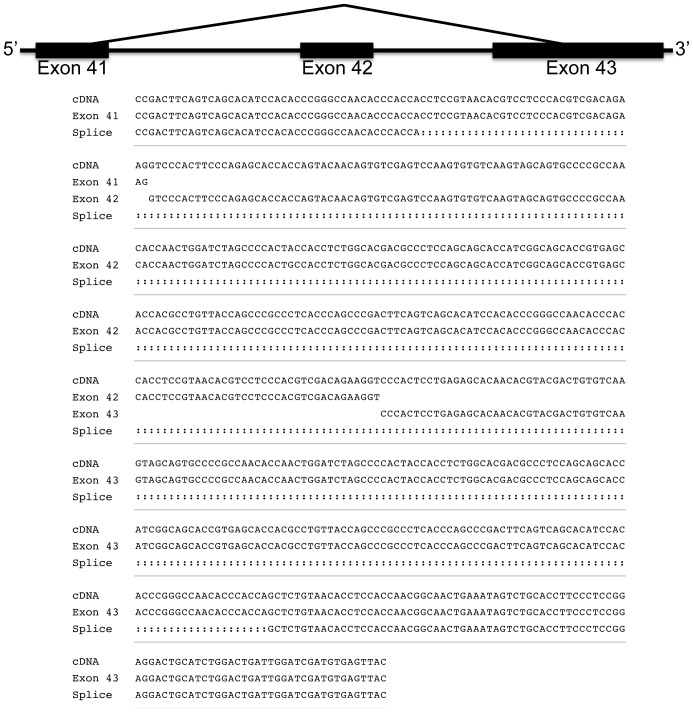
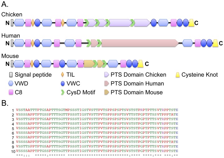
Intestinal mucin 2 (MUC2) encodes a heavily glycosylated, gel-forming mucin, which creates an important protective mucosal layer along the gastrointestinal tract in humans and other species. This first line of defense guards against attacks from microorganisms and is integral to the innate immune system. As a first step towards characterizing the innate immune response of MUC2 in different species, we report the cloning of a full-length, 11,359 bp chicken MUC2 cDNA, and describe the genomic organization and functional annotation of this complex, 74.5 kb locus. MUC2 contains 64 exons and demonstrates distinct spatiotemporal expression profiles throughout development in the gastrointestinal tract; expression increases with gestational age and from anterior to posterior along the gut. The chicken protein has a similar domain organization as the human orthologue, with a signal peptide and several von Willebrand domains in the N-terminus and the characteristic cystine knot at the C-terminus. The PTS domain of the chicken MUC2 protein spans ∼1600 amino acids and is interspersed with four CysD motifs. However, the PTS domain in the chicken diverges significantly from the human orthologue; although the chicken domain is shorter, the repetitive unit is 69 amino acids in length, which is three times longer than the human. The amino acid composition shows very little similarity to the human motif, which potentially contributes to differences in the innate immune response between species, as glycosylation across this rapidly evolving domain provides much of the musical barrier. Future studies of the function of MUC2 in the innate immune response system in chicken could provide an important model organism to increase our understanding of the biological significance of MUC2 in host defense and highlight the potential of the chicken for creating new immune-based therapies.


Conflict of interest statement

Competing Interests: The authors declare that Biomin Research Center, Tulln, Austria provided funding in terms of a research gift to TA at Purdue University. There are no restrictions on access to information, publication, or use of materials associated with this gift. Therefore, this partial funding by a commercial entity does not alter the authors adherence to all the PLOS ONE policies on sharing data and materials.

Figures

Figure 1
Figure 1. MUC2 cDNA contig.
A. MUC2 cDNA contigs. Sixteen overlapping RT-PCR amplicons and two RACE products are depicted. The double vertical lines represent the cDNA gap in the PTS domain. EST sequences are not shown. B. 5′ RACE. Representative image from 5′ RACE products size fractionated on a 1.2% 1× TAE gel. Molecular weight marker (Lane 1); 5′ RACE produce (Lane 2); Water control (Lane 3). C. 3′ RACE. Representative image from 3′ RACE products size fractionated on a 1.5% 1× TAE gel. Molecular weight marker (Lane 1); 3′ RACE produce (Lane 2); Water control (Lane 3). D. Genomic PCR. We amplified a 159 bp fragment that spanned the gap between exons 45 and 46. Exons are depicted to scale. The forward and reverse sequences from two spleen (Sp 1 and Sp2) samples are shown, along with the cDNA sequence for exons 45 and 46 and the primers. The last 9 bp from the reverse primer are not shown.
Figure 2
Figure 2. Northern blot analysis of intestinal MUC2.
A. Transcripts detected with a 5′ probe. B. Transcripts detected with a 3′ probe. Arrows show that both probes detected the same band. Membranes were stripped and demonstrated to show no signal prior to rehybridization. Lane M, ssRNA ladder (kilobase); Lane 1 and 2, 20 µg total RNA per lane. C. RNA integrity. Ethidium bromide-stained rRNAs of the same RNA samples electrophoresed on a 1.0% TAE gel are shown as an internal control. D. MUC2 expression following Eimeria infection. MUC2 transcripts detected in total RNA (30 µg) from cecal tonsils of chickens infected with Eimeria protozoa (+, lanes 3 and 4) or not (−, lane 1 and 2) were identified by probe 3 (1329-bp), which was synthesized by asymmetric PCR. Each lane represents pooled samples from 4 to 5 chickens. Arrow head: MUC2 mRNA.
Figure 3
Figure 3. Chicken MUC2 physical map and genomic organization.
A. Physical Map. The MUC2 locus lies on chromosome 5 between MUC5B and MUC6, and spans 74.5 kb of genomic DNA. B. Gene Structure. We identified an 11,359 bp MUC2 cDNA. Black vertical lines represent the location and relative size of the exons. There are two known assembly gaps in the chicken genome, based on the UCSC Nov 2011 Build (ICGSC Gallus_gallus-4.0/galGal4). Exon 36 (423 bp) is located in the smaller gap, and therefore not included in this figure, as it is unknown where it lies in relationship to its flanking exons. Exon 39 ends blindly in the large (∼21 kb) assembly gap. Exon 40 (29 bp, also not drawn in this map) is located within the gap, and exon 41 is intact on the other side of the gap. The location of these gaps are denoted by large black bars under the gene structure, and gaps are denoted in the genomic structure, when possible (the space between exons 33 and 35 is too small to insert a gap). Exons 34 (198 bp), 38 (44 bp) and 43 (869 bp), which are also not included in this figure) do not fall within the annotated gaps, but are missing in the genomic sequence, indicating errors in the assembly.
Figure 4
Figure 4. Functional Annotation of MUC2.
The compiled MUC2 cDNA is indicated by exon number (1–64). Black cells represent cDNAs, ESTs or predicted sequences that show the same exon structure as the reported cDNA; Grey boxes represent sequences that are shorter or longer than the compiled cDNA, or that contain significant numbers of mismatches compared to the intestinal MUC2 cDNA; blank cells indicate that this sequence is not present in the corresponding sequence. 1 G. gallus intestinal MUC2 cDNA from this report; 2Helmeted guineafowl; 3Turkey.
Figure 5
Figure 5. Expression of MUC2 in the gastrointestinal tract and brain.
RNAs were reverse transcribed using SMARTScribe™ (Clontech) with Oligo d(T) to generate long, full-length cDNA. We performed 33 cycles of RT-PCR amplification on 40 ng of cDNA with three sets of MUC2 primers. Alternating blank lanes lack reverse transcriptase. A. Exons 1–6. MUC2 is highly expressed in the proventriculus, duodenum, jejunum, ileum, colon, and cecal tonsil, with lower levels in the brain and minimal expression in the crop and ventriculus. Although these primers (P27 and P2) amplify genomic DNA, Genomic DNA controls demonstrate the lack of genomic contamination in all samples, indicating that observed expression is from cDNA B. Exons 16–23. MUC2 is highly expressed in the proventriculus, duodenum, jejunum, ileum, colon, and cecal tonsil, with lower levels in the brain and minimal expression in the crop and ventriculus. Although these primers (P7 and P8) amplify genomic DNA, Genomic DNA controls demonstrate the lack of genomic contamination in all samples, indicating that observed expression is from cDNA C Exons 44–65. A touchdown long-range PCR was used to amply the 3′ end of MUC2 using an internal primer and a primer targeting the exact end of the MUC2 cDNA (P30 and P29). This region demonstrates a very similar pattern of expression, with high levels detected in all tissues, except brain, which shows low-level expression,and ventriculus which has minimal expression. No expression is detected in the crop in this analysis. D. HPRT1 control gene. All samples express HPRT, and lack the presence of the genomic DNA band, indicating that the samples do not have genomic contamination. RT-PCR products were examined by electrophoresis through a 2.5% agarose gel in 0.5× TBE (A and B) or 1.2% TAE; water and genomic DNA were used as controls.
Figure 6
Figure 6. Temporal expression of MUC2 transcripts.
Quantification of MUC2 transcripts in embryonic (E) and post-hatch (H) ages in duodenum (top), jejunum (middle) and ileum (bottom) using qRT-PCR. MUC2 expression was normalized to 18S RNA. The MUC2 primers hybridize to exons 25 (P35) and 26 (P34) and generate a 135 bp product that spans intron 25 (1710 bp).
Figure 7
Figure 7. Alternate splicing.
We characterized one of the alternatively spliced products and determined that it was generated from an internal 3′ splice donor in exon 41. This transcript skipped exon 42, and used an alternate splice acceptor site in exon 43.
Figure 8
Figure 8. Cross-species comparison of the MUC2 protein structure.
A. Protein structure of chicken, human and mouse MUC2. InterProScan protein domain prediction analysis (www.ebi.ac.uk/Tools/pfa/iprscan/) indicates that the full-length chicken intestinal MUC2 transcript encodes a 3697 amino acid (aa) protein with a short signal peptide at the N-terminus, multiple von Willebrand factor domain structures (VWD, VWC), several cysteine-rich domains (C8), two trypsin Inhibitor-like cysteine-rich domains (TIL), a 1614 amino acid central PTS domain that is interspersed with four CysD motifs and a C-terminal cystine knot (CT). The structure of the human (5179 aa) and mouse (2680 aa) proteins shows strong homology on both sides of the central PTS domain. The two exceptions are that humans and mice lack the second TIL domain, and mice have an additional VWC motif. Although the N-terminal and C-terminal sequences are highly conserved amongst species, the PTS domain is highly divergent, containing different types and varying numbers of repeat cassettes within the central domain. In chicken, this region stretches between aa 1308 and 2922. The different colors in the cartoons represent the finding that the PTS domains are highly divergent among the three species. B. Sequence comparison of the 10 repeats within the PTS domain. Amino acids 1702 through 2763 demarcate this highly repetitive element, which spans exons 32 through 45. RADAR analysis (http://www.ebi.ac.uk/Tools/Radar/) indicates that these cassettes consist of three blocks of repetitive elements interspersed with two CysD domains. Each repeat is 69 amino acids in length and contains one of two short spacer motifs. Repeats 1 and 2 are located in block one, repeats 3–8 are located in block two and repeats 9 and 10 are located in block 3.

Similar articles

Cited by

References

    1. Hollingsworth MA, Swanson BJ (2004) Mucins in cancer: protection and control of the cell surface. Nat Rev Cancer 4: 45–60. - PubMed
    1. Hoorens PR, Rinaldi M, Li RW, Goddeeris B, Claerebout E, et al. (2011) Genome wide analysis of the bovine mucin genes and their gastrointestinal transcription profile. BMC Genomics 12: 140. - PMC - PubMed
    1. Lang T, Hansson GC, Samuelsson T (2007) Gel-forming mucins appeared early in metazoan evolution. Proc Natl Acad Sci U S A 104: 16209–16214. - PMC - PubMed
    1. McGuckin MA, Linden SK, Sutton P, Florin TH (2011) Mucin dynamics and enteric pathogens. Nat Rev Microbiology 9: 265–278. - PubMed
    1. Larsson JM, Karlsson H, Sjovall H, Hansson GC (2009) A complex, but uniform O-glycosylation of the human MUC2 mucin from colonic biopsies analyzed by nanoLC/MSn. Glycobiology 19: 756–766. - PubMed

Publication types

MeSH terms

Associated data