Expression and functional role of Sox9 in human epidermal keratinocytes
- PMID: 23349860
- PMCID: PMC3548846
- DOI: 10.1371/journal.pone.0054355
Expression and functional role of Sox9 in human epidermal keratinocytes
Abstract
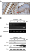
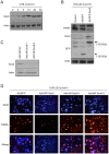
In this study, we investigated the expression and putative role of Sox9 in epidermal keratinocyte. Immunohistochemical staining showed that Sox9 is predominantly expressed in the basal layer of normal human skin epidermis, and highly expressed in several skin diseases including psoriasis, basal cell carcinoma, keratoacanthoma and squamous cell carcinoma. In calcium-induced keratinocyte differentiation model, the expression of Sox9 was decreased in a time dependent manner. When Sox9 was overexpressed using a recombinant adenovirus, cell growth was enhanced, while the expression of differentiation-related genes such as loricrin and involucrin was markedly decreased. Similarly, when rat skin was intradermally injected with the adenovirus expressing Sox9, the epidermis was thickened with increase of PCNA positive cells, while the epidermal differentiation was decreased. Finally, UVB irradiation induced Sox9 expression in cultured human epidermal keratinocytes, and keratinocytes are protected from UVB-induced apoptosis by Sox9 overexpression. Together, these results suggest that Sox9 is an important regulator of epidermal keratinocytes with putative pro-proliferation and/or pro-survival functions, and may be related to several cutaneous diseases that are characterized by abnormal differentiation and hyperproliferation.
Conflict of interest statement
Figures


Similar articles
-
Osteopontin facilitates ultraviolet B-induced squamous cell carcinoma development.J Dermatol Sci. 2014 Aug;75(2):121-32. doi: 10.1016/j.jdermsci.2014.05.002. Epub 2014 May 21. J Dermatol Sci. 2014. PMID: 24888687 Free PMC article.
-
Epidermal COX-2 induction following ultraviolet irradiation: suggested mechanism for the role of COX-2 inhibition in photoprotection.J Invest Dermatol. 2003 Oct;121(4):853-61. doi: 10.1046/j.1523-1747.2003.12495.x. J Invest Dermatol. 2003. PMID: 14632205
-
Epidermal expression of neuropilin 1 protects murine keratinocytes from UVB-induced apoptosis.PLoS One. 2012;7(12):e50944. doi: 10.1371/journal.pone.0050944. Epub 2012 Dec 10. PLoS One. 2012. PMID: 23251405 Free PMC article.
-
Effects of narrow band UVB (311 nm) irradiation on epidermal cells.Int J Mol Sci. 2013 Apr 17;14(4):8456-66. doi: 10.3390/ijms14048456. Int J Mol Sci. 2013. PMID: 23594996 Free PMC article. Review.
-
Calcium--a central regulator of keratinocyte differentiation in health and disease.Eur J Dermatol. 2014 Nov-Dec;24(6):650-61. doi: 10.1684/ejd.2014.2452. Eur J Dermatol. 2014. PMID: 25514792 Review.
Cited by
-
Cytoplasmic expression of SOX9 as a poor prognostic factor for oral squamous cell carcinoma.Oncol Rep. 2018 Nov;40(5):2487-2496. doi: 10.3892/or.2018.6665. Epub 2018 Aug 22. Oncol Rep. 2018. PMID: 30132562 Free PMC article.
-
Long-term expression pattern of melanocyte markers in light- and dark-pigmented dermo-epidermal cultured human skin substitutes.Pediatr Surg Int. 2015 Jan;31(1):69-76. doi: 10.1007/s00383-014-3622-7. Epub 2014 Oct 18. Pediatr Surg Int. 2015. PMID: 25326121
-
Sox9 expression in canine epithelial skin tumors.Eur J Histochem. 2015 Jul 9;59(3):2514. doi: 10.4081/ejh.2015.2514. Eur J Histochem. 2015. PMID: 26428883 Free PMC article.
-
Effect of Adalimumab on Gene Expression Profiles of Psoriatic Skin and Blood.J Drugs Dermatol. 2016 Aug 1;15(8):988-94. J Drugs Dermatol. 2016. PMID: 27538000 Free PMC article.
-
SOX9: a stem cell transcriptional regulator of secreted niche signaling factors.Genes Dev. 2014 Feb 15;28(4):328-41. doi: 10.1101/gad.233247.113. Genes Dev. 2014. PMID: 24532713 Free PMC article.
References
-
- Kalinin AE, Kajava AV, Steinert PM (2002) Epithelial barrier function: assembly and structural features of the cornified cell envelope. Bioessays 24: 789–800. - PubMed
-
- Hennings H, Michael D, Cheng C, Steinert P, Holbrook K, et al. (1980) Calcium regulation of growth and differentiation of mouse epidermal cells in culture. Cell 19: 245–254. - PubMed
-
- Piao MS, Choi JY, Lee DH, Yun SJ, Lee JB, et al. (2011) Differentiation-dependent expression of NADP(H):quinone oxidoreductase-1 via NF-E2 related factor-2 activation in human epidermal keratinocytes. J Dermatol Sci 62: 147–153. - PubMed
Publication types
MeSH terms
Substances
LinkOut - more resources
Full Text Sources
Other Literature Sources
Medical
Research Materials
Miscellaneous

