. 2013;8(1):e54366.
doi: 10.1371/journal.pone.0054366. Epub 2013 Jan 18.

Separate developmental programs for HLA-A and -B cell surface expression during differentiation from embryonic stem cells to lymphocytes, adipocytes and osteoblasts

Affiliations

Separate developmental programs for HLA-A and -B cell surface expression during differentiation from embryonic stem cells to lymphocytes, adipocytes and osteoblasts

Hardee J Sabir et al. PLoS One. 2013.

Abstract

A major problem of allogeneic stem cell therapy is immunologically mediated graft rejection. HLA class I A, B, and Cw antigens are crucial factors, but little is known of their respective expression on stem cells and their progenies. We have recently shown that locus-specific expression (HLA-A, but not -B) is seen on some multipotent stem cells, and this raises the question how this is in other stem cells and how it changes during differentiation. In this study, we have used flow cytometry to investigate the cell surface expression of HLA-A and -B on human embryonic stem cells (hESC), human hematopoietic stem cells (hHSC), human mesenchymal stem cells (hMSC) and their fully-differentiated progenies such as lymphocytes, adipocytes and osteoblasts. hESC showed extremely low levels of HLA-A and no -B. In contrast, multipotent hMSC and hHSC generally expressed higher levels of HLA-A and clearly HLA-B though at lower levels. IFNγ induced HLA-A to very high levels on both hESC and hMSC and HLA-B on hMSC. Even on hESC, a low expression of HLA-B was achieved. Differentiation of hMSC to osteoblasts downregulated HLA-A expression (P = 0.017). Interestingly HLA class I on T lymphocytes differed between different compartments. Mature bone marrow CD4(+) and CD8(+) T cells expressed similar HLA-A and -B levels as hHSC, while in the peripheral blood they expressed significantly more HLA-B7 (P = 0.0007 and P = 0.004 for CD4(+) and CD8(+) T cells, respectively). Thus different HLA loci are differentially regulated during differentiation of stem cells.


Conflict of interest statement

Competing Interests: The authors have declared that no competing interests exist.

Figures

Figure 1
Figure 1. A schematic diagram representing the order of differentiation of the cells used in this study.
Figure 2
Figure 2. Expression of HLA-A and HLA-B in hESC and hMSC.
Representative flow histograms showing HLA-A and -B expression in stem cell lines using allele-specific antibodies (anti-A2, -A3, -B7, and -B13). (A) In separate experiments, hESC lines (huES9 and KMEB2) showed a low expression of HLA-A alleles (A2 and A3, respectively) and no expression of the HLA-B alleles (B13 and B7, respectively). IFNγ stimulation up-regulated HLA-A alleles and induced of modest expression of HLA-B alleles. (B) Panel B shows representative flow histograms of an hMSC cell line (ToB11-13) both un-stimulated and IFNγ stimulated (72 hr) demonstrating a high constitutive expression of HLA-A2 and a relatively low expression of HLA-B7. Both antigens were up-regulated after stimulation with IFNγ. Panel (C) compares the expression of HLA-A and HLA-B between hESC (huES9) and hMSC (ToB11-13) during basal, un-induced conditions and after stimulation for 72 h with 25 ng/µl IFNγ. HLA expression was measured by flowcytometry as molecules of equivalent fluorochromes (MEF).
Figure 3
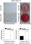
Figure 3. Differentiation of hMSC to adipocytes and osteoblasts.
Representative images for Oil O Red and Alizarin red staining demonstrating adipocyte and osteoblasts differentiation of hMSC cell lines cultured under adipogenic and osteogenic conditions, respectively. Non-differentiated cells were not stained (A and D), while the adipocyte differentiated cells were stained with Oil O red (B) and Alizarin red stained the matrix formed by osteoblasts (E). qPCR data shows marked up-regulation of PPAR gamma gene expression after adipocyte differentiation (C) and up-regulation of Collagen 1 gene expression in osteoblast differentiated cells (F).
Figure 4
Figure 4. Expression of HLA-A and HLA-B on hMSC and in their differentiated adipocyte and osteoblast progenies.
Locus-specific HLA antigen expression before and after 13 days of differentiation of hMSC to adipocytes (A), or 16 days of differentiation to osteoblasts (B) in one hMSC-Tert4 cell line and three primary hMSC cell lines (Table 1). The analyzed HLA-A alleles were HLA-A2 (N = 3) and -A3 (N = 2). The analyzed HLA-B alleles were HLA-B7 (N = 3), -B27 (N = 1) and -B8 (N = 1) (data pooled according to locus, each data point represents mean value for 3 experiments for each cell line).
Figure 5
Figure 5. Cell surface HLA expression of hHSC and lymphocytes derived from BM and peripheral blood.
All allelic forms of HLA studied were highly expressed in all the cell types shown. However, HLA-A2 was expressed at significantly higher levels than HLA-B7 and -B8 on hHSC, BM CD4+ T cells, and BM CD8+ T cells. When BM lymphocytes and peripheral blood lymphocytes were compared, there was a marginal (but non-significant) difference in the expression of HLA-A2 and -B8 alleles which tended to have higher expression in peripheral blood than in BM. A significantly higher expression level of HLA-B7 was observed on peripheral blood CD4+ and CD8+ T lymphocytes. In peripheral blood, the expression of HLA-B8 was significantly lower than HLA-A2 and -B7 on CD4+ and CD8+ T lymphocytes. Peripheral CD19+ lymphocytes expressed similar amounts of HLA-A2, -B7 and -B8.

Similar articles

Cited by

References

    1. Abbas AK, Lichtman AH, Pillai S (2010) Cellular and Molecular Immunology. Saunders/Elsevier. 566 p.
    1. Roitt IM, Brostoff J, Male DK (2001) Immunology. Mosby. 480 p.
    1. Parham P (2009) The Immune System. Garland Science. 506 p.
    1. Ahmed-Ansari A, Tadros TS, Knopf WD, Murphy DA, Hertzler G, et al. (1988) Major histocompatibility complex class I and class II expression by myocytes in cardiac biopsies posttransplantation. Transplantation 45: 972–978. - PubMed
    1. Apps R, Murphy SP, Fernando R, Gardner L, Ahad T, et al. (2009) Human leucocyte antigen (HLA) expression of primary trophoblast cells and placental cell lines, determined using single antigen beads to characterize allotype specificities of anti-HLA antibodies. Immunology 127: 26–39. - PMC - PubMed

Publication types

MeSH terms