. 2013;8(1):e54382.
doi: 10.1371/journal.pone.0054382. Epub 2013 Jan 18.

CK2 phosphorylates Sec31 and regulates ER-To-Golgi trafficking

Affiliations

CK2 phosphorylates Sec31 and regulates ER-To-Golgi trafficking

Mayuko Koreishi et al. PLoS One. 2013.

Abstract

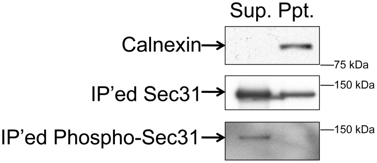
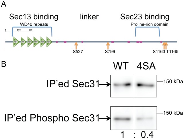
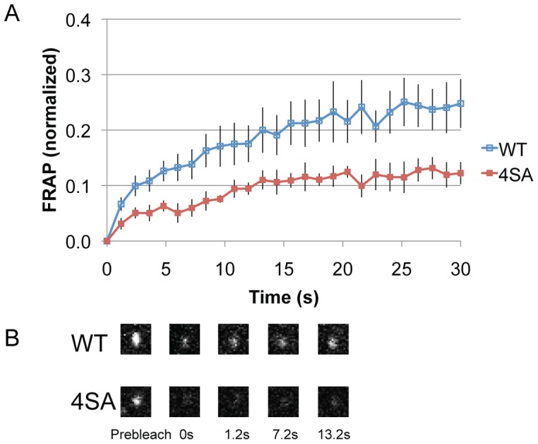
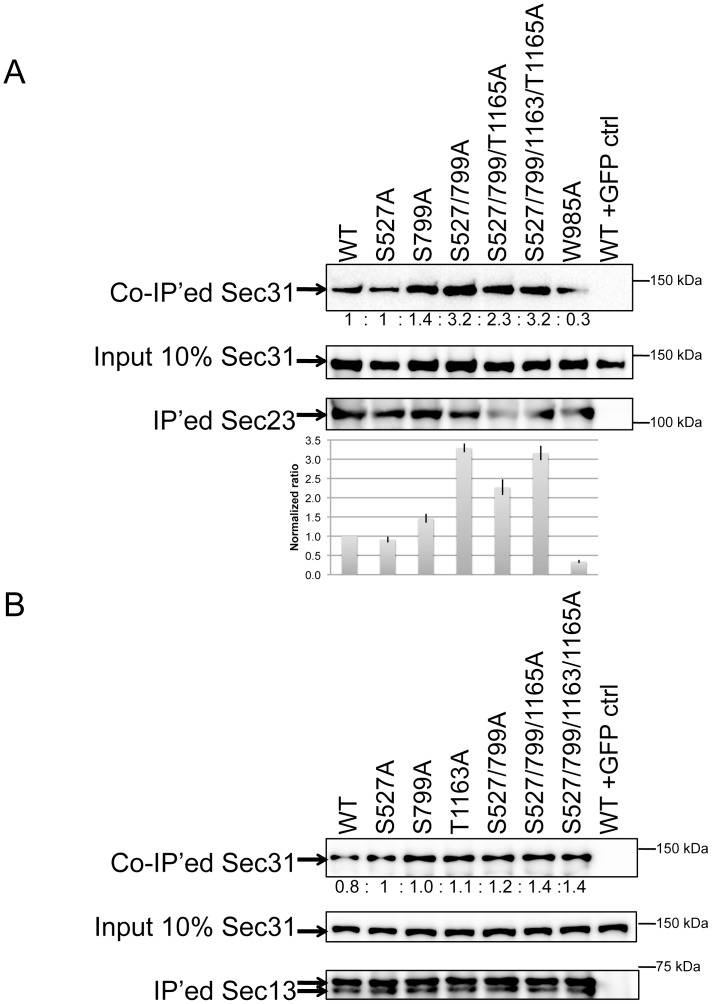
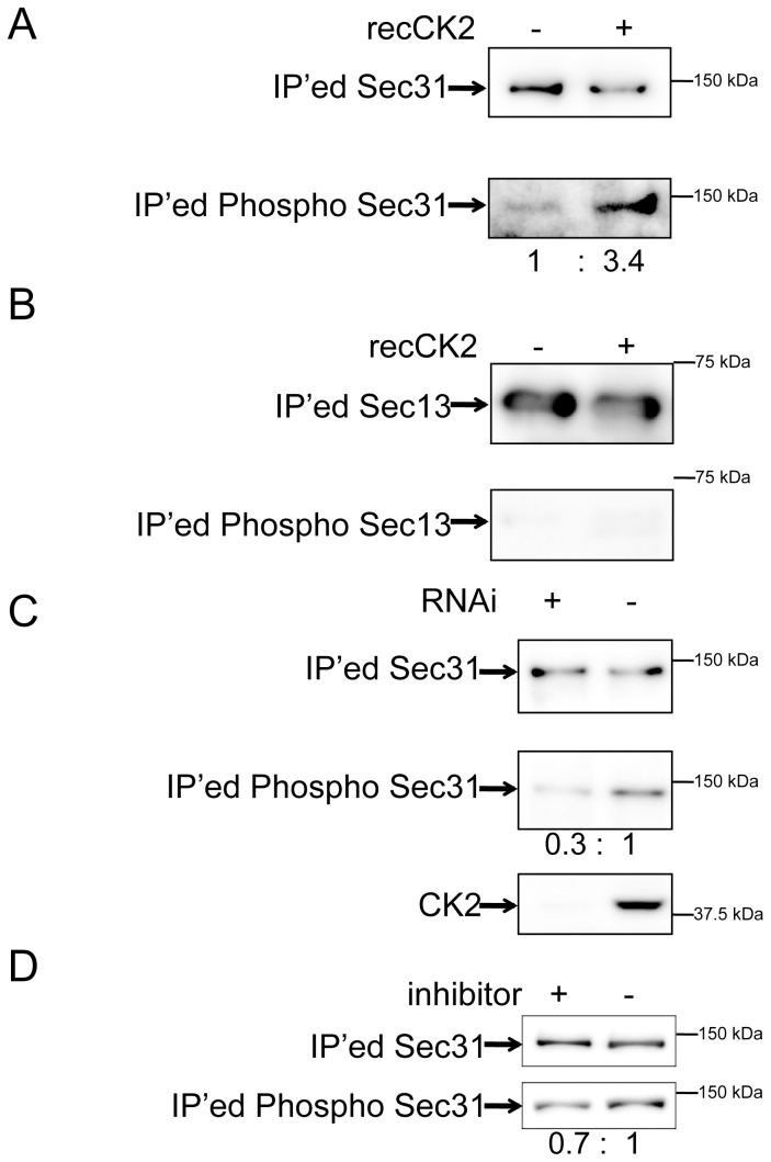
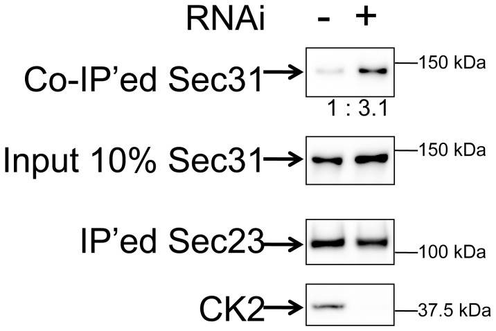
Protein export from the endoplasmic reticulum (ER) is an initial and rate-limiting step of molecular trafficking and secretion. This is mediated by coat protein II (COPII)-coated vesicles, whose formation requires small GTPase Sar1 and 6 Sec proteins including Sec23 and Sec31. Sec31 is a component of the outer layer of COPII coat and has been identified as a phosphoprotein. The initiation and promotion of COPII vesicle formation is regulated by Sar1; however, the mechanism regulating the completion of COPII vesicle formation followed by vesicle release is largely unknown. Hypothesizing that the Sec31 phosphorylation may be such a mechanism, we identified phosphorylation sites in the middle linker region of Sec31. Sec31 phosphorylation appeared to decrease its association with ER membranes and Sec23. Non-phosphorylatable mutant of Sec31 stayed longer at ER exit sites and bound more strongly to Sec23. We also found that CK2 is one of the kinases responsible for Sec31 phosphorylation because CK2 knockdown decreased Sec31 phosphorylation, whereas CK2 overexpression increased Sec31 phosphorylation. Furthermore, CK2 knockdown increased affinity of Sec31 for Sec23 and inhibited ER-to-Golgi trafficking. These results suggest that Sec31 phosphorylation by CK2 controls the duration of COPII vesicle formation, which regulates ER-to-Golgi trafficking.


Conflict of interest statement

Competing Interests: The authors have declared that no competing interests exist.

Figures

Figure 1
Figure 1. Membrane associated Sec31 is not phosphorylated.
HeLa cells were subjected to subcellular fractionations to prepare cytosol (Sup) and membranes (Ppt) by ultracentrifugation. The recovery of the transmembrane protein, calnexin in the membrane fraction (Ppt) indicates that the fractionation was performed properly. Subsequently, Sec31 from both fractions were immunoprecipitated and subjected to western blotting with anti-Sec31 and anti-phospho-serine/threonine antibodies.
Figure 2
Figure 2. Identification of the phosphorylation sites in Sec31.
(A) The predicted domain structure of human Sec31 by SMART (http://smart.embl-heidelberg.de). The domain structure of Sec31 is: N-terminal WD repeats for Sec13 binding, a linker region, and a C-terminal extensin-like proline-rich domain for Sec23 binding. The phosphorylation sites identified by mass-spectrometry were indicated by arrows. (B) HEK293 cells were transfected with FLAG-tagged wild type (WT) Sec31 or its S527A/S799A/S1163A/T1165A mutant (4SA). Tagged proteins were immunoprecipitated with antibodies to the FLAG tag and subjected to western blotting with anti-FLAG and anti-phospho-serine/threonine antibodies. The immunoprecipitated (IP’ed) phospho-Sec31 levels were normalized with IP’ed Sec31 levels and expressed as the normalized ratio.
Figure 3
Figure 3. The dynamic of membrane association and dissociation of Sec31 is reduced with the non-phosphorylatable mutant.
(A) Shown are the results of fluorescence recovery after photobleaching (FRAP) of Sec31 and its 4SA. GFP-tagged wild type Sec31 (WT) and the 4SA mutant were expressed transiently in HeLa cells. A GFP positive dot was photobleached and subsequent fluorescence recovery was monitored for 30 s with 1.2 s intervals. Eight dots were photobleached per cell and results were normalized. Represented are the average of ∼7 experiments. Bars, SEM. (B) The representative images at the indicated time points of Figure 3A. Sizes, 1.85 (w) x 1.85 (h) µm.
Figure 4
Figure 4. Dephosphorylation of Sec31 at Serines 527 and 799 increases its affinity to Sec23.
(A) HEK293 cells were co-transfected with indicated FLAG-tagged Sec31 mutants and GFP-tagged Sec23. Sec23 was immunoprecipitated with anti-GFP beads and subjected to western blotting with anti-FLAG and anti-GFP antibodies. The “input 10% Sec31” is a western blot of 10% aliquots of total cell lysates. The normalized ratio of Sec31 bound to Sec23 was shown below. W985A is a known Sec23-binding defect mutant . The most right lane is a negative control with GFP. The graph below the blots shows the average of the quantification of three independent experiments with SD. (B) FLAG-Sec31 and GFP-Sec13 (instead of GFP-Sec23) were co-expressed and their interactions detected by co-immunoprecipitation and western blotting following the procedures described in Figure 4A. The lane on right most is a negative control with GFP.
Figure 5
Figure 5. CK2 phosphorylates human Sec31.
(A) FLAG-Sec31 expressed in HRK293 cells was immunoprecipitated and incubated with/without recombinant CK2 (recCK2) followed by western blotting with anti-phospho serine/threonine and anti-FLAG antibodies (IP’ed Sec31). The phospho-Sec31 levels were normalized to IP’ed Sec31 levels and expressed as the normalized ratio. (B) FLAG-Sec13 expressed in HRK293 cells was immunoprecipitated, treated and subjected to western blotting as described in (A). (C) Cells were first transfected with siRNA1 to CK2 alpha1 (RNAi +) or to eGFP (RNAi −). After 2 days incubation, cells were transfected with FLAG-tagged Sec31 transfection followed by immunoprecipitation and western blotting as described in (A). Total cell lysates were also analyzed for CK2 by western blotting to determine the depletion efficiency as shown in the bottom panel. (D) FLAG-Sec31 expressed in HRK293 cells treated with or without CK2 inhibitor was immunoprecipitated and subjected to western blotting as described in (A).
Figure 6
Figure 6. Depletion of CK2 increases Sec31’s affinity to Sec23.
Two days before the FLAG-tagged Sec31 and GFP-Sec23 co-transfection, HEK293 cells were transfected with (RNAi +) or without siRNA1 to CK2 alpha1 (RNAi −). GFP-tagged Sec23 was immunoprecipitated with anti-GFP beads and subjected to western blot with anti-FLAG and anti-GFP antibodies. The “input 10% Sec31” is a western blot of 10% aliquots of total cell lysates in order to show the transfection and binding efficiency of Sec31. The co-immunoprecipitated (co-IP’ed) Sec31 were normalized with input Sec31 and expressed as the normalized ratio. In order to check the efficiency of CK2 RNAi, western blotting of the total cell lysates with anti-CK2 antibodies was shown in the bottom panel.
Figure 7
Figure 7. Depletion of CK2 reduces ER to Golgi trafficking.
(A) HeLa cells stably expressing secretory alkaline phosphatase (SEAP) were transfected with siRNA1 to CK2 alpha1 (RNAi, 20 nM) or to eGFP (mock). After 90 h, cells were washed and fed with new media. In 6 h, aliquots of culture supernatants were collected and their phosphatase activities were measured in triplicates. Data are presented as a secretion index, which is the ratio of SEAP activity detected in the culture medium to the cellular SEAP activity. Bars, SD (n = 3). (B) CK2 alpha1 siRNA1- or mock-treated cells were transfected with VSV-G-tsO45-YFP plasmid and incubated at the restrictive temperature of 40oC overnight before shifting them to the permissive temperature of 32°C. In 15 min, cells were fixed, permeabilized, and labeled with anti-GM130 antibodies to define the Golgi region. Cell images were captured and analyzed using Image J and Photoshop (n = ∼20 cells). Error bars represent the SEM of three independent experiments. (C) The representative images at the indicated time points of Figure 7B. Bar, 10 µm. (D) HeLa cells stably expressing secretory alkaline phosphatase (SEAP) were transfected with wild type (WT), 4SA mutant (4SA) or 4SD mutant (4SD). After 24 h, cells were washed and fed with new media with or without CK2 inhibitor 1 (Calbiochem). In 6 h, aliquots of culture supernatants were collected and their phosphatase activities were measured in triplicates. Data are presented as a normalized secretion (Secretion index of WT = 100%). Bars, SD (n = 3).

References

    1. Lee MC, Orci L, Hamamoto S, Futai E, Ravazzola M, et al. (2005) Sar1p N-terminal helix initiates membrane curvature and completes the fission of a COPII vesicle. Cell 122: 605–617. - PubMed
    1. Sato K, Nakano A (2005) Dissection of COPII subunit-cargo assembly and disassembly kinetics during Sar1p-GTP hydrolysis. Nat Struct Mol Biol 12: 167–174. - PubMed
    1. Weissman JT, Plutner H, Balch WE (2001) The mammalian guanine nucleotide exchange factor mSec12 is essential for activation of the Sar1 GTPase directing endoplasmic reticulum export. Traffic 2: 465–475. - PubMed
    1. Futai E, Hamamoto S, Orci L, Schekman R (2004) GTP/GDP exchange by Sec12p enables COPII vesicle bud formation on synthetic liposomes. EMBO J 23: 4146–4155. - PMC - PubMed
    1. Barlowe C (2002) Three-dimensional structure of a COPII prebudding complex. Dev Cell 3: 467–468. - PubMed

Publication types