. 2013;8(1):e54384.
doi: 10.1371/journal.pone.0054384. Epub 2013 Jan 22.

In vitro secondary structure of the genomic RNA of satellite tobacco mosaic virus

Affiliations

In vitro secondary structure of the genomic RNA of satellite tobacco mosaic virus

Shreyas S Athavale et al. PLoS One. 2013.

Abstract

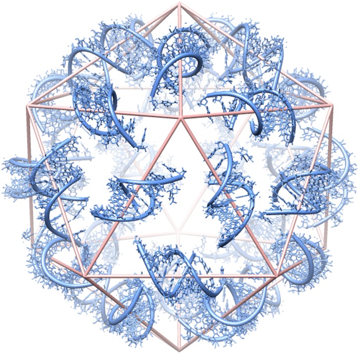
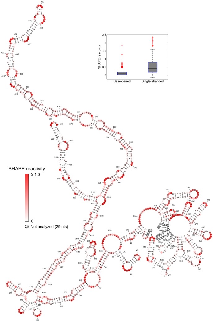
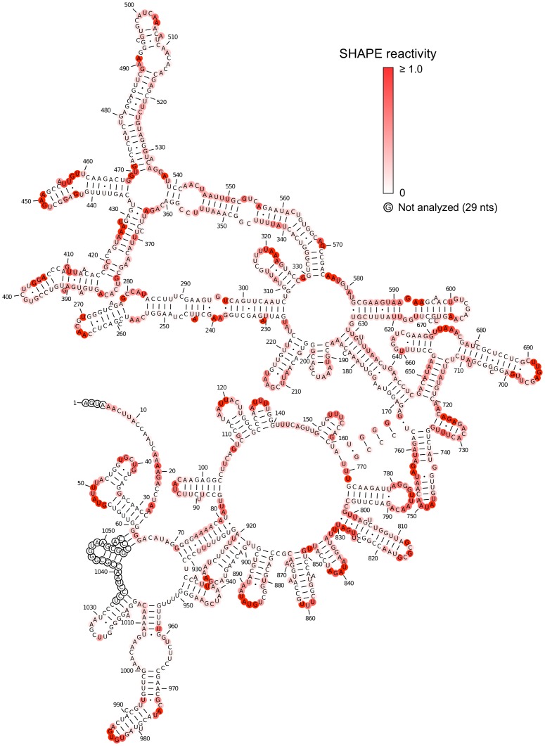
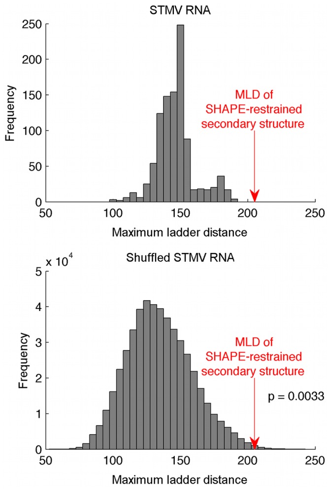
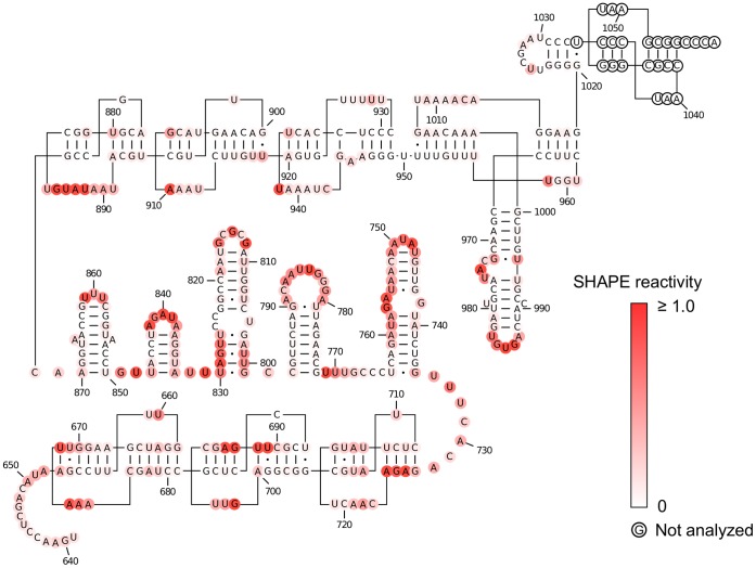
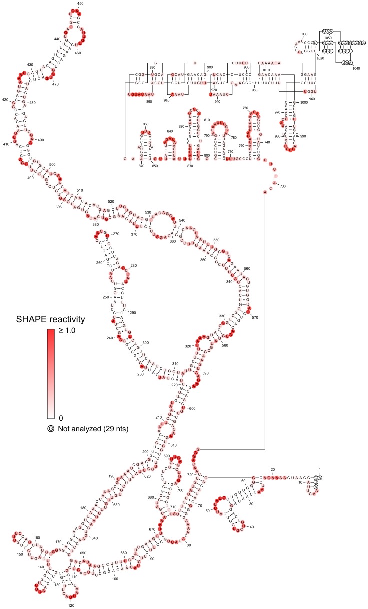
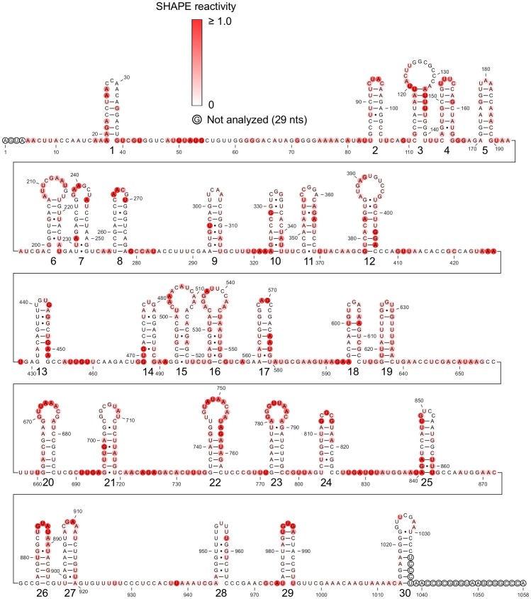
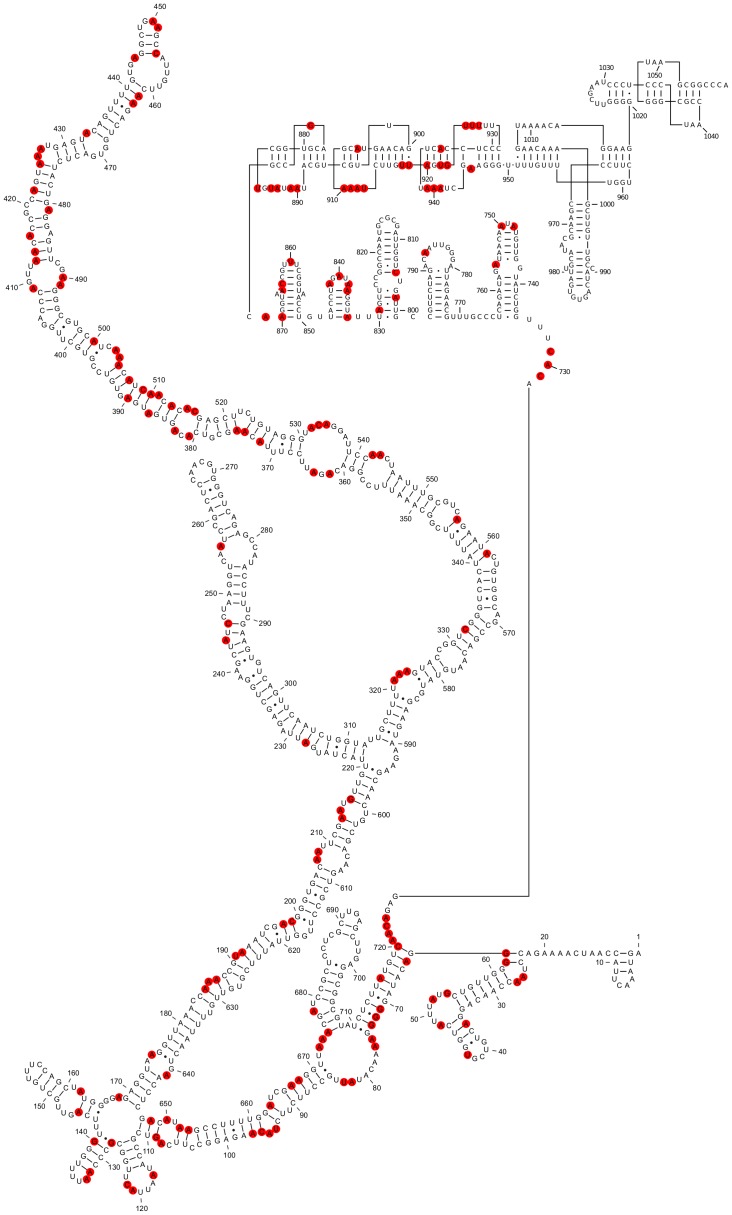
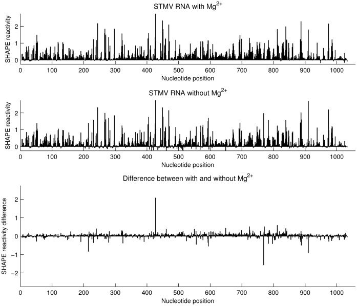
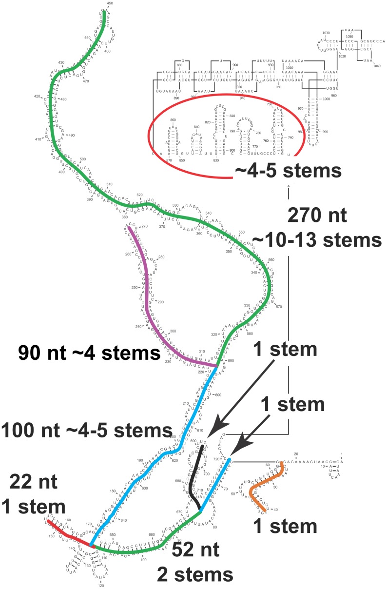
Satellite tobacco mosaic virus (STMV) is a T = 1 icosahedral virus with a single-stranded RNA genome. It is widely accepted that the RNA genome plays an important structural role during assembly of the STMV virion. While the encapsidated form of the RNA has been extensively studied, less is known about the structure of the free RNA, aside from a purported tRNA-like structure at the 3' end. Here we use selective 2'-hydroxyl acylation analyzed by primer extension (SHAPE) analysis to examine the secondary structure of in vitro transcribed STMV RNA. The predicted secondary structure is unusual in the sense that it is highly extended, which could be significant for protecting the RNA from degradation. The SHAPE data are also consistent with the previously predicted tRNA-like fold at the 3' end of the molecule, which is also known to hinder degradation. Our data are not consistent with the secondary structure proposed for the encapsidated RNA by Schroeder et al., suggesting that, if the Schroeder structure is correct, either the RNA is packaged as it emerges from the replication complex, or the RNA undergoes extensive refolding upon encapsidation. We also consider the alternative, i.e., that the structure of the encapsidated STMV RNA might be the same as the in vitro structure presented here, and we examine how this structure might be organized in the virus. This possibility is not rigorously ruled out by the available data, so it remains open to examination by experiment.


Conflict of interest statement

Competing Interests: The authors have declared that no competing interests exist.

Figures

Figure 1
Figure 1. Distribution of double-helical RNA segments in the STMV virion.
The crystal structure of STMV reveals 30 segments of double-helical RNA (blue). Each helix contains 9 base pairs, centered on a crystallographic two-fold axis. An icosahedral cage (pink) is shown for reference. Adopted from .
Figure 2
Figure 2. SHAPE-restrained secondary structure model for free STMV RNA.
Nucleotides are colored according to their SHAPE reactivity (see scale). Inset shows a box plot comparing the distribution of SHAPE reactivity values between base paired and single-stranded nucleotides. Each grey box represents the interquartile range (IQR) of the data; the bottom and top edges of the box are the 25th and 75th percentiles, respectively. The band near the middle of each box is the median value. The whiskers above and below each box extend to the most extreme data points not considered outliers. Outliers are plotted individually as crosses. Points are outliers if they are greater than 1.5 IQR from the 75th percentile or less than 1.5 IQR from the 25th percentile. In this secondary structure model, the distribution for base paired nucleotides is narrower and has a much lower median value than the distribution for single-stranded nucleotides.
Figure 3
Figure 3. Minimum free energy (MFE) structure obtained for STMV RNA without the SHAPE data.
The structure was predicted using RNAstructure with default parameters. Nucleotides are colored according to their SHAPE reactivity (see scale). The SHAPE data are not consistent with this model, since several base paired regions have high reactivity values.
Figure 4
Figure 4. Histogram of maximum ladder distance values calculated for STMV RNA and shuffled STMV RNA sequences.
The MLD of the SHAPE-restrained structure is much higher than the MLDs of 1000 suboptimal structures predicted for the STMV RNA sequence (top). The extreme MLD of the SHAPE-restrained structure is unlikely to have occurred by chance: the bottom histogram was obtained using 1000 suboptimal structures for each of 500 randomly shuffled sequences with the same length and nucleotide composition as STMV. Fewer than 0.4% of these structures have MLDs greater than the MLD of the SHAPE-restrained STMV structure.
Figure 5
Figure 5. Predicted secondary structure at the 3′ end of STMV RNA.
Secondary structure for the 406 3′-terminal nucleotides of STMV RNA proposed by Gultyaev et al. . Nucleotides are colored according to their SHAPE reactivity (see scale). The SHAPE data supports the tRNA-like structure and the five stem-loops (nucleotides 728–1058), but does not support the second pseudoknot domain (nucleotides 653–727).
Figure 6
Figure 6. SHAPE-restrained secondary structure of free STMV RNA with a tRNA-like fold at the 3′ end.
This alternate model of the STMV RNA was obtained by combining the SHAPE-restrained secondary structure predicted separately for nucleotides 1–727 (Figure 2) with the Gultyaev et al. prediction for nucleotides 728–1058 (Figure 5). Nucleotides are colored according to their SHAPE reactivity (see scale). The extended central domain (nucleotides 64–720) is identical to that of Figure 2.
Figure 7
Figure 7. Schroeder secondary structure model for encapsidated STMV RNA.
Schroeder et al. predicted this secondary structure on the basis of the co-replicational folding and assembly hypothesis, along with chemical probing data . Nucleotides are colored according to their SHAPE reactivity (see scale), and the hairpin loops are numbered from 1 to 30. Hairpins 1, 3, 10–13, 17, 21–22, and 25 are clearly inconsistent with the SHAPE data.
Figure 8
Figure 8. Mapping the chemical probing data from Schroeder et al. onto the SHAPE-restrained secondary structure of in vitro transcribed STMV RNA.
Red circles indicate nucleotides modified by DMS, kethoxal, or CMCT. The data do not appear to clearly rule out the proposed secondary structure of residues 1–730. A substantial number of the modifications occur in predicted loops, bulges, and single-stranded regions (67 out of 119 hits). Many of the reactive base-paired nucleotides are in A-U or G-U base pairs immediately adjacent to a predicted bulge loop (e.g., 128, 185, 187, 192, 213, 413–414, 556, 561, 652–653, 663, 675), while others (382–390 and 503–515) are in a predicted stem that has two bulges and has no run of more than three consecutive base pairs, so it should be prone to fraying.
Figure 9
Figure 9. Effect of Mg2+ on the SHAPE reactivity profile of free STMV RNA.
SHAPE reactivities for STMV RNA in the presence (top) and absence (middle) of Mg2+. The difference plot (bottom) shows that 10 mM Mg2+ has little effect on the SHAPE reactivity profile.
Figure 10
Figure 10. Identification of possible double-helical stems corresponding to those seen in the crystal structure.
There are 30 stems in the crystal structure, each containing nine base pairs with an additional base stacked at each 3′ end, i.e., 20 nucleotides (Figure 1). A model that connects successive stems would require something on the order of 5–10 nucleotides per connection. This figure shows how our secondary structure model might be organized to fit into the STMV capsid, with a sufficient number of stems to cover the 30 edges of the icosahedral frame, as required by Figure 1.

References

    1. Dodds JA (1998) Satellite tobacco mosaic virus. Annual Review of Phytopathology 36: 295–310. - PubMed
    1. Schneemann A (2006) The structural and functional role of RNA in icosahedral virus assembly. Annu Rev Microbiol 60: 51–67. - PubMed
    1. Gossele V, Fache I, Meulewaeter F, Cornelissen M, Metzlaff M (2002) SVISS - a novel transient gene silencing system for gene function discovery and validation in tobacco plants. Plant Journal 32: 859–866. - PubMed
    1. Larson SB, Day J, Greenwood A, McPherson A (1998) Refined structure of satellite tobacco mosaic virus at 1.8 Å resolution. J Mol Biol 277: 37–59. - PubMed
    1. Larson SB, McPherson A (2001) Satellite tobacco mosaic virus RNA: Structure and implications for assembly. Curr Opinion Struct Biol 11: 59–65. - PubMed

Publication types

LinkOut - more resources