. 2013;8(1):e54410.
doi: 10.1371/journal.pone.0054410. Epub 2013 Jan 22.

Requirement for Dlgh-1 in planar cell polarity and skeletogenesis during vertebrate development

Affiliations

Requirement for Dlgh-1 in planar cell polarity and skeletogenesis during vertebrate development

Charlene Rivera et al. PLoS One. 2013.

Abstract

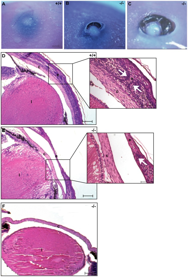
The development of specialized organs is tightly linked to the regulation of cell growth, orientation, migration and adhesion during embryogenesis. In addition, the directed movements of cells and their orientation within the plane of a tissue, termed planar cell polarity (PCP), appear to be crucial for the proper formation of the body plan. In Drosophila embryogenesis, Discs large (dlg) plays a critical role in apical-basal cell polarity, cell adhesion and cell proliferation. Craniofacial defects in mice carrying an insertional mutation in Dlgh-1 suggest that Dlgh-1 is required for vertebrate development. To determine what roles Dlgh-1 plays in vertebrate development, we generated mice carrying a null mutation in Dlgh-1. We found that deletion of Dlgh-1 caused open eyelids, open neural tube, and misorientation of cochlear hair cell stereociliary bundles, indicative of defects in planar cell polarity (PCP). Deletion of Dlgh-1 also caused skeletal defects throughout the embryo. These findings identify novel roles for Dlgh-1 in vertebrates that differ from its well-characterized roles in invertebrates and suggest that the Dlgh-1 null mouse may be a useful animal model to study certain human congenital birth defects.


Conflict of interest statement

Competing Interests: The authors have declared that no competing interests exist.

Figures

Figure 1
Figure 1. Generation of Dlgh-1 null mice.
(A) Schematic representation of the targeting of the Dlgh-1 allele. WT, wild-type allele; C, construct, plasmid containing loxP sites flanking exon 8 of Dlgh-1 and neo cassette, and the TK cassette; F, the floxed allele allele, containing lox P sites flanking Dlgh-1′s exon 8 and the neo cassette; R, recombined allele, desired recombination event lacking exon 8 and neo. Abbreviations: 7, exon 7; 8, exon 8; 9, exon 9; P, PstI; triangles, lox P sequences; neo, neomycin; TK, thymidine kinase. 3′ probe for Southern blot analysis is denoted by black bars. Fragments expected from PstI digestion and 3′ probe hybridization are depicted by double-arrowed lines. (B) Southern blot analysis of PstI digested genomic DNA isolated from mice that were WT (+/+), heterozygote (+/−), or null (−/−) for the Dlgh-1 allele. Sizes of hybridizing bands are shown in kb. (C) Western blot analysis of Dlgh-1 protein levels. Protein lysates of brain, heart, lung, kidney, skin, eye, liver, and mouse embryo fibroblasts (MEF) from Dlgh-1 wild type (+/+), heterozygote (+/−), or null (−/−) mice were resolved by SDS-PAGE and immunoblotted with an anti-Dlgh-1 antibody, as described in Materials and Methods. Membranes were reprobed with an anti-β-actin antibody as a loading control.
Figure 2
Figure 2. Phenotype of Dlgh-1−/− mice.
(A) Newborn Dlgh-1−/− mice are smaller then their Dlgh-1+/+ counterparts and have a shortened snout and mandible (arrows). (B–C) Hematoxylin and eosin stained paraffin sections of FVB/NJ E14.5 Dlgh-1+/+ (B) and Dlgh-1−/− (C) mice. At this developmental stage, the reduced size of the Dlgh-1−/− is noticeable. (D–E). Dissected heads of Dlgh-1+/+ and Dlgh-1−/− C57BL/6J E18.5 embryos showing the cleft palate (asterisk*) in Dlgh-1−/− mice. (F–G) Frontal sections of Dlgh-1+/+ (F) and Dlgh-1−/− (G) E18.5 embryos on the C57BL/6J background showing the absence of the nasopharyngeal passage (NP, arrow in F). (H–I) Sagittal H&E stained sections of E18.5 Dlgh-1+/+ (H) and Dlgh-1−/− (I) embryos on the FVB/NJ background showing the dome-shaped head of Dlgh-1−/− mice, the absence of the nasopharyngeal passage (NP), and the shortened snout and mandible (arrows). NC, nasal cavity; NP, nasopharyngeal passage; P, pharynx; T, tongue. Scale bar = 250 µm.
Figure 3
Figure 3. Dlgh-1−/− mice display defects in eyelid closure.
E18.5 Dlgh-+/+− and Dlgh-1−/− mice on the C57BL/6 background were collected and analyzed for the presence of closed eyelids. Dlgh-1+/+ mice (A) form closed eyelids while Dlgh-1−/− (B, C) littermates show defects in eyelid closure. (D–F) Hematoxylin and eosin staining of histological sections collected from E18.5 mice show completely closed eyelids in Dlgh-1+/+ mice (D) whereas eyelids in Dlgh-1−/− mice were only partially closed (E) or completely open (F). Insets show a higher magnification of the eyelids. Arrows show the position of the tips of the dermis in Dlgh-1+/+ (D) and Dlgh-1−/− (E) embryos, which failed to extend fully in the Dlgh-1−/− eye. Scale bar = 250 µm and 150 µm for insets. c, cornea; e, eyelids; l, lens.
Figure 4
Figure 4. Expression of Dlgh-1 and Vangl2 proteins in the eyelid of Dlgh-1+/+ and Dlgh-1−/− mice.
Sections of sagittally oriented, paraffin embedded heads from newborn Dlgh-1+/+ (A–C) and Dlgh-1−/− (D–F) mice on the C57BL/6J background were subjected to double immunofluorescence analysis with anti-Dlgh-1 (green) and anti-Vangl2 (red) antibodies. Shown are representative merged and unmerged images of the staining in the eyelid of Dlgh-1+/+ (A–C) and Dlgh-1−/− (D–F) eyelids. Dlgh-1 (A, B) and Vangl2 (A, C) localized to the membranes of the epithelium (arrow in A) including the site of eyelid fusion, in Dlgh-1+/+ mice. Staining for Dlgh-1 was absent in eyelid of the Dlgh-1−/− mice (E) and Vangl2 accumulated in the cytoplasm rather than in the membrane (D, F). e, epithelium; ep, epidermis; f, eyelid fusion. Scale bar = 50 µm.
Figure 5
Figure 5. A fraction of Dlgh-1−/− mice display defects in neural tube closure.
E16.5 Dlgh-1+/+ and Dlgh-1−/− embryos on the C57BL/6J background were collected, examined for overt defects, and photographed under a dissecting microscope. Profile (A) and dorsal (B) views of an E16.5 Dlgh-1 +/+ embryo. Profile (C) and dorsal (D) views of an E16.5 Dlgh-1−/− embryo. In addition to craniorachischisis (C, D arrows), the Dlgh-1−/− embryo exhibits gastroschisis (C, arrowhead) and a rightward kink to the body axis (D).
Figure 6
Figure 6. Dlgh-1−/− mice display defects in outer cell hair polarity.
(A, B) Representative images of the OHC and IHC layers in the cochlea of Dlgh-1+/+ and Dlgh-1−/− mice. The organ of Corti was isolated from the cochleae of E18.5 Dlgh-1+/+ (A) and Dlgh-1 −/− (B) embryos, stained with phalloidin to visualize the actin bundles of the inner and outer hair cells, and viewed by confocal microscopy. Arrows indicate cells with misoriented stereociliary bundles in OHC3 row of the Dlgh-1−/− mice. (C) Measurement of misorientation of sterociliary bundles in OHC3 layer of Dgh-1+/+ and Dlgh-1−/− mice. The angle of sterociliary bundles in OHC3 of Dlgh-1+/+ and Dlgh-1−/− mice was measured in reference to a line parallel to the neural-abneural axis and perpendicular to the plane of the pillar cells. Plotted is the deviation in degrees from the neural-abneural axis for each OHC3 bundle measured in (4 cochleae from 3 mice of each genotype, 36 cells for Dlgh-1+/+ and 33 cells for Dlgh-1−/− mice). No deviation from the neural-abneural axis was assigned a value of 0°. The average angle deviation from 0° for OHC3 bundles in Dlgh-1−/− cochlea (33.7°+/−1.869, red bar) was significantly higher (p = 1.5×10⁻⁹) than that of the Dlgh-1+/+ (17.3°+/−0.869, red bar). (D, E) Immunohistochemical detection of Dlgh-1 protein in the cochlea of E15.5 mouse embryos. Transverse sections of paraffin embedded heads from control Dlgh-1+/+ mice were subjected to immunohistochemistry using (D) an anti-Dlgh-1 specific antibody and alkaline phosphatase detection or (E) no primary antibody. The pink color in the epithelium of the cochlear duct and cartilage in (D) is indicative of the presence of Dlgh-1 protein in these structures. (F) Immunofluorescent detection of Dlgh-1 protein in the cochlear hair cells of P2 mice control. Fixed cochlear explants from Dlgh-1+/+ mice were subjected to immunofluorescence using an anti-Dlgh-1 (green) antibody. Arrows show concentration of Dlgh-1 on the basal-lateral membranes of the OHC cells. IHC, inner hair cells, OHC1, 2, 3, outer hair cell rows 1, 2, and 3. c, cartilage in cochlea, d, cochlear duct. Scale bar = 50 µm.
Figure 7
Figure 7. Dlgh-1−/− mice display defects in skeletogenesis.
Eviscerated carcasses of E18.5 Dlgh-1+/+, Dlgh-1+/−, and Dlgh-1−/− mice on the FVB/NJ background were prepared and stained for cartilage and bone with alcian blue and alizarin red, respectively. Craniofacial skeleton in (A) Dlgh-1+/+, (B) Dlgh-1+/−, and (C) Dlgh-1−/− mice. Arrows show loss of bone formation in Dlgh-1+/− and Dlgh-1−/− mice. F, frontal bone; IP, intraparietal bone; P, parietal bone; N, nasal; Ro, rostral bone; RTP, retrotemporal process; T, temporal bone. (D–G) Axial structures of Dlgh-1+/+ (D) and Dlgh-1−/− mice (E). Forelimb (F) and hindlimb (G) structures. Arrows show the absence of bone formation in Dlgh-1−/− mice. S, sternebrae; H, humerus; R, radius; U, ulna; F, femur; Fi, fibula; T, tibia. Scale bar in A-E = 1000 µm, in F = 650 µm and in G = 500 µm.

Similar articles

Cited by

References

    1. Woods DF, Hough C, Peel D, Callaini G, Bryant PJ (1996) Dlg protein is required for junction structure, cell polarity, and proliferation control in Drosophila epithelia. J Cell Biol 134: 1469–1482. - PMC - PubMed
    1. Bilder D, Li M, Perrimon N (2000) Cooperative regulation of cell polarity and growth by Drosophila tumor suppressors. Science 289: 113–116. - PubMed
    1. Wang Y, Nathans J (2007) Tissue/planar cell polarity in vertebrates: new insights and new questions. Development 134: 647–658. - PubMed
    1. Montcouquiol M, Rachel RA, Lanford PJ, Copeland NG, Jenkins NA, et al. (2003) Identification of Vangl2 and Scrb1 as planar polarity genes in mammals. Nature 423: 173–177. - PubMed
    1. Bossinger O, Klebes A, Segbert C, Theres C, Knust E (2001) Zonula adherens formation in Caenorhabditis elegans requires dlg-1, the homologue of the Drosophila gene discs large. Dev Biol 230: 29–42. - PubMed

Publication types

MeSH terms

LinkOut - more resources