. 2013;8(1):e54426.
doi: 10.1371/journal.pone.0054426. Epub 2013 Jan 17.

Differential effects of rapamycin and dexamethasone in mouse models of established allergic asthma

Affiliations

Differential effects of rapamycin and dexamethasone in mouse models of established allergic asthma

Elizabeth M Mushaben et al. PLoS One. 2013.

Abstract

The mammalian target of rapamycin (mTOR) plays an important role in cell growth/differentiation, integrating environmental cues, and regulating immune responses. Our lab previously demonstrated that inhibition of mTOR with rapamycin prevented house dust mite (HDM)-induced allergic asthma in mice. Here, we utilized two treatment protocols to investigate whether rapamycin, compared to the steroid, dexamethasone, could inhibit allergic responses during the later stages of the disease process, namely allergen re-exposure and/or during progression of chronic allergic disease. In protocol 1, BALB/c mice were sensitized to HDM (three i.p. injections) and administered two intranasal HDM exposures. After 6 weeks of rest/recovery, mice were re-exposed to HDM while being treated with rapamycin or dexamethasone. In protocol 2, mice were exposed to HDM for 3 or 6 weeks and treated with rapamycin or dexamethasone during weeks 4-6. Characteristic features of allergic asthma, including IgE, goblet cells, airway hyperreactivity (AHR), inflammatory cells, cytokines/chemokines, and T cell responses were assessed. In protocol 1, both rapamycin and dexamethasone suppressed goblet cells and total CD4(+) T cells including activated, effector, and regulatory T cells in the lung tissue, with no effect on AHR or total inflammatory cell numbers in the bronchoalveolar lavage fluid. Rapamycin also suppressed IgE, although IL-4 and eotaxin 1 levels were augmented. In protocol 2, both drugs suppressed total CD4(+) T cells, including activated, effector, and regulatory T cells and IgE levels. IL-4, eotaxin, and inflammatory cell numbers were increased after rapamycin and no effect on AHR was observed. Dexamethasone suppressed inflammatory cell numbers, especially eosinophils, but had limited effects on AHR. We conclude that while mTOR signaling is critical during the early phases of allergic asthma, its role is much more limited once disease is established.


Conflict of interest statement

Competing Interests: The authors have declared that no competing interests exist.

Figures

Figure 1
Figure 1. Study protocols.
A: Protocol 1: Re-Exposure Study: Mice were sensitized to HDM by 3 i.p. injections followed by 2 intranasal HDM or saline (control) exposures. One group of mice (group 1) was sacrificed after the first round of allergen exposures while the other two groups rested/recovered for 6 weeks. After 6 weeks of rest, group 2 was sacrificed before allergen re-exposure. Mice in group 3 were re-exposed to intranasal HDM or saline (control) twice and were treated with rapamycin or dexamethasone during this exposure period. Mice were sacrificed 48 hours after the last HDM exposure. B: Protocol 2: Reversal Study: Mice were exposed to HDM intranasally 3 times per week for 3 or 6 weeks. Starting at week 4, groups of mice were treated with rapamycin or dexamethasone by i.p. injection six days per week for the remaining three weeks. Mice were sacrificed 48 hours after the last HDM exposure.
Figure 2
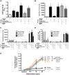
Figure 2. Protocol 1- Allergic sensitization, inflammatory cell numbers in the BALF, and AHR after HDM re-exposure.
A, HDM-specific IgE levels were increased in HDM exposed (group 1) and HDM re-exposed (group 3) mice compared to saline controls. Rapamycin (Rapa) attenuated HDM-induced increases in IgE after allergen re-exposure, while dexamethasone (Dex) had no effect (n = 4–12 mice/group). *p<0.05 versus saline; <p<0.05 versus HDM Rest; ∧p<0.05 versus HDM re-exposure; #p<0.05 versus Dex. B, Total BALF cell numbers were increased in HDM exposed mice in groups 1 and 3, but not in mice that rested for 6 weeks after HDM exposure (group 2) and unaltered by Rapa or Dex treatment (n = 4–12 mice/group). *p<0.05 versus saline; <p<0.05 versus HDM Rest. C, Total numbers of macrophages and eosinophils were increased after HDM re-exposure (group 3), but not in HDM rest (group 2) animals in the BALF. Total neutrophil numbers in the BALF were slightly increased after HDM re-exposure in Rapa treated mice. Rapa did not suppress HDM-induced increases in eosinophils. Eosinophil numbers were lower in Dex treated mice compared to HDM re-exposed and Rapa treated groups, but still higher then saline control (n = 4–12 mice/group). *p<0.05 versus saline; ∧p<0.05 versus HDM re-exposure; <p<0.05 versus HDM Rest; #p<0.05 versus Dex. D, The percentage of eosinophils in the BALF was increased in all HDM exposed groups except HDM rest mice, while the percentage of macrophages were reduced. (n = 4–12 mice/group). *p<0.05 versus saline; ∧p<0.05 versus HDM re-exposure; <p<0.05 versus HDM Rest; #p<0.05 versus Dex. E, AHR was increased after the initial set of HDM exposures (group 1) compared to saline controls and was still increased after allergen re-exposure (group 3). Neither Rapa nor Dex suppressed HDM-induced increases in AHR after allergen re-exposure. AHR was similar to controls in HDM rest mice (group 2) (n = 6–14 mice/group). *p<0.05 versus saline.
Figure 3
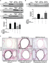
Figure 3. Protocol 1- Goblet cell markers in HDM re-exposed mice.
A, Western blot analysis of lung homogenates showed that the goblet cell protein, CLCA3, was increased after the initial set of HDM exposures (group 1) and after HDM re-exposure (group 3). Both rapamycin (Rapa) and dexamethasone (Dex) treatment attenuated HDM-induced increases in CLCA3 (n = 3–5 mice/group). *p<0.05 versus saline; <p<0.05 versus HDM Rest; ∧p<0.05 versus HDM re-exposed; #p<0.05 versus Dex. B, HDM-induced increases in the transcription factor, SPDEF, were suppressed by Rapa, but not Dex after allergen re-exposure (group 3). (n = 3 mice/group). *p<0.05 versus saline; ∧p<0.05 versus HDM re-exposed; #p<0.05 versus Dex. C, Muc5AC immunostaining was increased in lung epithelial cells after HDM exposure (group 1) compared to saline controls, but was attenuated after 6 weeks of rest (group 2). After HDM re-exposure (Group 3), Muc5AC staining was increased again compared to saline controls and HDM rest (group 2). Rapamycin reduced Muc5AC staining in the lung, but staining was still elevated compared to saline controls. Dexamethasone treatment appeared to have minimal effects on HDM-induced increases in Muc5AC staining.
Figure 5
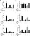
Figure 5. Protocol 1- Th1, Th2, and Th17 cytokines and chemokines in BALF of HDM re-exposed mice.
A, Levels of INF-γ appeared to be lower after rapamycin (Rapa) treatment in HDM re-exposed mice, but were not statistically different between any of the groups (n = 3–7 mice/group). B, IL-17A levels in the BALF were not different between groups (n = 3–8 mice/group). C, IL-13 levels were increased after the initial set of HDM exposures (group 1), but not after allergen re-exposure (group 3). Neither Rapa nor dexamethasone (Dex) had any effect on IL-13 levels after HDM re-exposure (n = 3–8 mice/group). D, IL-4 levels were increased after the first set of HDM exposures (group 1), but not after HDM re-exposure (group 3). Rapa treatment augmented IL-4 levels after HDM re-exposure, but Dex did not. E, IL-5 levels were increased in group 1 after the initial set of HDM exposures, but not after HDM re-exposure (group 3). IL-5 levels were unaffected by Rapa or Dex treatment following HDM re-exposure (n = 3–8 mice/group). *p<0.05 versus saline. F, Eotaxin 1 levels were increased in group 1 after HDM exposure. After HDM re-exposure, Rapa augmented eotaxin 1 levels (n = 3–8 mice/group). *p<0.05 versus saline; <p<0.05 versus HDM Rest; ∧p<0.05 versus HDM re-exposed; #p<0.05 versus Dex.
Figure 4
Figure 4. Protocol 1- T cell populations in mice after HDM re-exposure.
A, HDM-induced increases in total CD4+ lung cells after allergen re-exposure were suppressed by rapamycin (Rapa) and dexamethasone (Dex) (n = 4–12 mice/group). *p<0.05 versus saline; ∧p<0.05 versus HDM re-exposed. B, Activated T cells (CD69+Foxp3) were increased after HDM re-exposure and suppressed by Rapa and Dex (n = 4–12 mice/group). *p<0.05 versus saline; ∧p<0.05 versus HDM re-exposed; #p<0.05 versus dex. C, CD69+Foxp3 activated T cells, as a percentage of total CD4+ T cells was also increased after HDM re-exposure and suppressed by Rapa, but not Dex (n = 4–12 mice/group) *p<0.05 versus saline; ∧p<0.05 versus HDM re-exposed; #p<0.05 versus dex. D, Lung effector T cells (CD44+Foxp3) were increased after HDM re-exposure and attenuated by Rapa and Dex (n = 4–12 mice/group). *p<0.05 versus saline; ∧p<0.05 versus HDM re-exposed. E, CD44+Foxp3 effector cells, as a percentage of total CD4+ T cells were increased in all HDM re-exposed groups and not suppressed by Rapa or Dex (n = 4–12 mice/group). *p<0.05 versus saline. F, Total lung regulatory T cells (Foxp3+CD25+) were increased after HDM re-exposure and suppressed by Rapa and Dex (n = 4–12 mice/group). *p<0.05 versus saline; ∧p<0.05 versus HDM re-exposed. G, Foxp3+CD25+cells, as a percentage of total lung CD4+ T cells, were slightly reduced by Rapa treatment, but not Dex (n = 4–12 mice/group). ∧p<0.05 versus HDM re-exposed. H, The ratio of Foxp3+CD25+ regulatory T cells/CD44+Foxp3 effector T cells was lower in HDM re-exposed mice compared to saline controls, as well as Rapa and Dex groups (n = 4–12 mice/group). *p<0.05 versus saline.
Figure 7
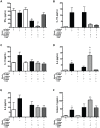
Figure 7. Protocol 2- T cell populations after chronic HDM exposure.
A, Total CD4+ T cells were increased after 6 weeks of HDM and suppressed by rapamycin (Rapa) and dexamethasone (Dex) (n = 4–12 mice/group). *p<0.05 versus saline; ∧p<0.05 versus HDM. B, Total activated T cells (CD69+Foxp3) were increased in mice after 6 weeks of chronic HDM exposure and suppressed by Rapa and Dex (n = 6–8 mice/group). *p<0.05 versus saline; ∧p<0.05 versus HDM. C, CD69+Foxp3 activated T cells, when assessed as a percentage of total CD4+ T cells, were increased after HDM exposure and unaffected by Rapa and Dex (n = 6–8 mice/group). *p<0.05 versus saline. D, Total effector T cells (CD44+Foxp3) were increased after 6 weeks of HDM exposure and suppressed by Rapa and Dex (n = 6–8 mice/group). *p<0.05 versus saline; ∧p<0.05 versus HDM. E, CD44+Foxp3 effector T cells, when expressed as a percentage of total CD4+ T cells, were increased after HDM exposure and attenuated by Rapa and Dex (n = 6–8 mice/group). *p<0.05 versus saline; ∧p<0.05 versus HDM. F, Total lung regulatory T cells (Foxp3+CD25+) were increased after chronic HDM exposure and suppressed by Rapa and Dex (n = 6–8 mice/group). *p<0.05 versus saline; ∧p<0.05 versus HDM. G, Foxp3+CD25+ T cells, as a percentage of total lung CD4+ T cells, were reduced by Dex, but not by Rapa (n = 6–8 mice/group). ∧p<0.05 versus HDM; # p<0.05 versus Rapa. H, The ratio of regulatory T cells Foxp3+CD25+ to CD44+Foxp3 effector T cells was decreased in HDM exposed mice compared to saline controls (n = 6–8 mice/group). *p<0.05 versus saline.
Figure 6
Figure 6. Protocol 1- Western blot of P-S6 and P-Akt in lung homogenates of HDM re-exposed mice.
A, P-S6, a downstream mediator of mTOR complex 1 signaling, was increased in HDM re-exposed mice (group 3) and this was blocked by rapamycin (Rapa) treatment (n = 3–5 mice/group). *p<0.05 versus saline; ∧p<0.05 versus HDM re-exposed. B, P-Akt, a downstream mediator of mTOR complex 2 signaling, was increased after allergen re-exposure in Rapa treated mice compared to saline controls (n = 3–5 mice/group), but unaffected by Dex. *p<0.05 versus saline; #p<0.05 versus Rapa.
Figure 8
Figure 8. Protocol 2- Th1, Th2, and Th17 cytokines and chemokines in the BALF after chronic HDM exposure.
A, INF-γ was suppressed after 6 weeks of HDM exposure and in the rapamycin (Rapa) treated HDM group (n = 3–5 mice/group). *p<0.05 versus saline; # p<0.05 versus Dex. B, No significant differences in IL-17A levels were observed between animal groups, although there were trends for increased IL-17 in the HDM exposed group and lower leves in mice exposed to HDM and treated with Rapa or dexamethasone (Dex) during weeks 4–6 (n = 3–5 mice/group). C, IL-13 levels were not significantly altered with HDM exposure, by Rapa or by Dex treatment in this model (n = 3–5 mice/group). D, IL-4 levels were increased after 3 weeks of HDM compared to saline controls. After 6 weeks of HDM, IL-4 levels were higher in the Rapa treated group. IL-4 levels were similar between HDM and Dex treated mice (n = 3–5 mice/group). *p<0.05 versus saline; ∧p<0.05 versus HDM; # p<0.05 versus Dex. E, No statistically significant differences in IL-5 levels were observed between any group of mice (n = 3–5 mice/group). F, Eotaxin 1 levels were increased after 3 weeks of HDM exposure and were higher in the Rapa treated group after 6 weeks of chronic HDM compared to saline controls (n = 3–5 mice/group). *p<0.05 versus saline.

References

    1. Bateman ED, Hurd SS, Barnes PJ, Bousquet J, Drazen JM, et al. (2008) Global strategy for asthma management and prevention: GINA executive summary. The Eur Respir J 31: 143–178. - PubMed
    1. Hamid Q, Tulic M (2009) Immunobiology of asthma. Annu Rev Physiol 71: 489–507. - PubMed
    1. Holgate ST (2012) Innate and adaptive immune responses in asthma. Nat Med 18: 673–683. - PubMed
    1. Kim HY, DeKruyff RH, Umetsu DT (2010) The many paths to asthma: phenotype shaped by innate and adaptive immunity. Nature immunology 11: 577–584. - PMC - PubMed
    1. Hesselmar B, Aberg B, Eriksson B, Aberg N (2000) Asthma in children: prevalence, treatment, and sensitization. Pediatr Allergy Immunol 11: 74–79. - PubMed

Publication types

MeSH terms