. 2013;8(1):e54454.
doi: 10.1371/journal.pone.0054454. Epub 2013 Jan 22.

Sod1 deficiency reduces incubation time in mouse models of prion disease

Affiliations

Sod1 deficiency reduces incubation time in mouse models of prion disease

Shaheen Akhtar et al. PLoS One. 2013.

Abstract

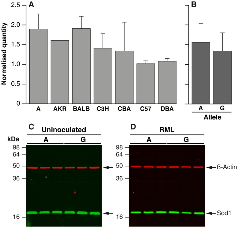
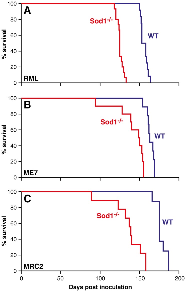
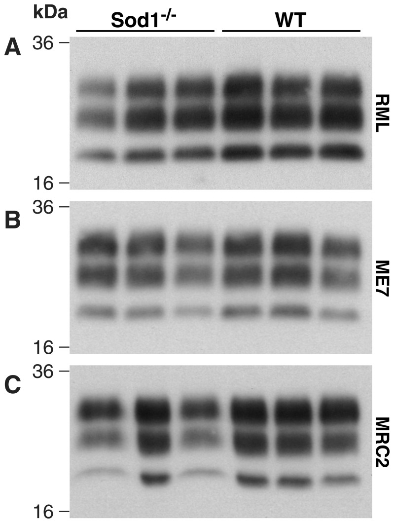
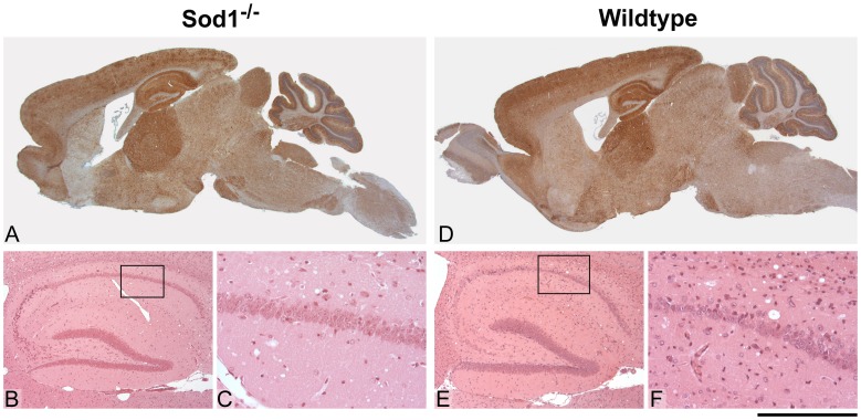
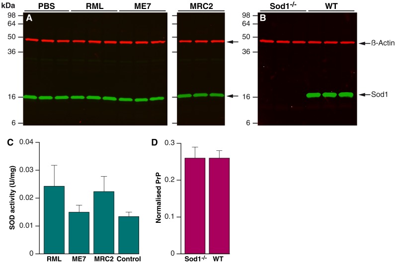
Prion infections, causing neurodegenerative conditions such as Creutzfeldt-Jakob disease and kuru in humans, scrapie in sheep and BSE in cattle are characterised by prolonged and variable incubation periods that are faithfully reproduced in mouse models. Incubation time is partly determined by genetic factors including polymorphisms in the prion protein gene. Quantitative trait loci studies in mice and human genome-wide association studies have confirmed that multiple genes are involved. Candidate gene approaches have also been used and identified App, Il1-r1 and Sod1 as affecting incubation times. In this study we looked for an association between App, Il1-r1 and Sod1 representative SNPs and prion disease incubation time in the Northport heterogeneous stock of mice inoculated with the Chandler/RML prion strain. No association was seen with App, however, significant associations were seen with Il1-r1 (P = 0.02) and Sod1 (P<0.0001) suggesting that polymorphisms at these loci contribute to the natural variation observed in incubation time. Furthermore, following challenge with Chandler/RML, ME7 and MRC2 prion strains, Sod1 deficient mice showed highly significant reductions in incubation time of 20, 13 and 24%, respectively. No differences were detected in Sod1 expression or activity. Our data confirm the protective role of endogenous Sod1 in prion disease.


Conflict of interest statement

Competing Interests: JC is a director and shareholder of D-Gen limited, an academic spin-out company in the field of prion diagnosis, decontamination and therapeutics. This does not alter the authors’ adherence to all the PLOS One policies on sharing data and materials.

Figures

Figure 1
Figure 1. Sod1 expression in mouse brain.
Panel A and B. Quantification of Sod1 mRNA expression in whole brain by real-time RT-PCR. N = 6 for all groups and samples were run in triplicate. All samples were duplexed for Sod1 (Fam-label) and an endogenous control GAPDH, β-actin or Thy-1 (Vic-label). Expression level is expressed in arbitrary units as normalised by the geometric mean of the quantity of the endogenous controls (y-axis). Error bars represent the standard deviation. (A) Sod1 mRNA expression level for parental strain of the HS mice (except LP). (B) Sod1 mRNA expression level grouped by allele (A/G) (A = A, BALB, C3H, C57; G = AKR, CBA, DBA). No significant difference was observed between the groups. Panels (C) and (D). Quantification of Sod1 protein in whole brain (10% homogenate, weight/volume) from n = 3 A allele mice (C57Bl/6) and G allele mice (FVB/N). FVB/N mice were used to represent a G allele strain rather than an HS parental strain due to availability of tissue. Samples were immunoblotted with rabbit polyclonal anti-human SOD1(Abcam) and detected with IRDye800CW (green) conjugated goat anti-rabbit IgG (Li-Cor). Anti-β-actin mouse monoclonal antibody (Sigma) was also included as a loading control and was detected using IRDye680 (red) conjugated goat anti-mouse IgG (Li-Cor). Fluorescence was visualised using an Odyssey infra-red imager (Li-Cor). (C) Uninoculated mice, (D) Terminally sick Chandler/RML prion inoculated mice.
Figure 2
Figure 2. Kaplan-Meier survival curves.
Data are shown as % of animals (female) surviving (y-axis) plotted against the number of days post inoculation (x-axis). (A) Transmission of Chandler/RML prion strain to Sod1−/− and Sod1+/+ (WT) litter mate controls (B) Transmission of ME7 prion strain to Sod1−/− and Sod1+/+ (WT). (C) Transmission of MRC2 mouse adapted BSE prion strain to Sod1−/− and Sod1+/+ (WT). A reduction in mean incubation time of 20%, 13%, and 24% was seen in A-C respectively. This reduction in survival was statistically significant for each transmission (P<0.001, Kaplan-Meier log-rank survival test).
Figure 3
Figure 3. Western blots of PrPSc from infected mouse brains.
10% w/v brain homogenates (n = 3 per group) were digested with proteinase K and immunoblotted with anti-PrP monoclonal antibody ICSM35 (D-Gen Ltd, UK). (A) Transmission of Chandler/RML prions to Sod1−/− and Sod1+/+ (WT) litter mate controls. (B) Transmission of ME7 prion strain to Sod1−/− and Sod1+/+ (WT) litter mate controls. (C) Transmission of MRC2 mouse adapted BSE prion strain to Sod1−/− and Sod1+/+ (WT) litter mate controls. No differences were seen between the two groups regardless of prion strain.
Figure 4
Figure 4. RML histology.
Histological features of Chandler/RML prion transmission to Sod1−/− (A–C) and wild type control (D–F) mice. Panels A and D show distribution of disease-associated PrP by immunohistochemistry using anti-PrP monoclonal antibody ICSM35. Panels B, C, E and F show detail from the hippocampus and are stained with haematoxylin and eosin (H&E) to visualise spongiform change and neuronal loss. There is almost no neuronal loss in the Sod1−/− mice but mild neuronal loss is seen in the wild type animals. Overall, the pattern of spongiosis, gliosis and PrP distribution are similar between the two groups, however, the distribution of disease-associated PrP is patchier in the knockouts especially in the cortex. Scale bar corresponds to 3 mm (A, D), 660 µm (B, E) or 160 µm (C, F).
Figure 5
Figure 5. Quantification of Sod and PrPC
. 10% weight/volume brain homogenates immunoblotted with rabbit polyclonal anti-human SOD1 (Abcam) and detected with IRDye800CW (green) conjugated goat anti-rabbit IgG (Li-Cor). Anti-β-actin mouse monoclonal antibody (Sigma) was also included as a loading control and was detected using IRDye680 (red) conjugated goat anti-mouse IgG (Li-Cor). Fluorescence was visualised using an Odyssey infra-red imager (Li-Cor). (A) Brains from Sod1+/+ wild type mice inoculated with PBS compared with end stage Sod1+/+ wild type mice inoculated with RML, ME7 and MRC2 prion strains. No differences were seen between the groups. (B) Uninfected mice. (C) Total SOD enzymatic activity in 10% (w/v) Sod1+/+ (WT) brains. Brains from terminally sick mice infected with RML, ME7 and MRC2 were compared with uninfected mice. Samples were run in triplicate with n = 6 for each group. Data are shown normalised by total protein content (µg/ml) as determined by a Bradford protein assay (mean ± standard deviation). No significant difference was seen between the groups. (D) PrPc levels in Sod1−/− and Sod1+/+ (WT) litter mate control mice by ELISA. PrPc levels (µg/ml) were determined in triplicate using 10% (weight/volume) brain homogenate for Sod1−/− (n = 3) and Sod1+/+ (n = 3) in a PrP specific ELISA . Data are shown normalised by total protein content (µg/ml and x 1000) as determined by a BCA assay (mean ± standard deviation). No significant difference was seen between the two groups.

Similar articles

Cited by

References

    1. Collinge J (2001) Prion diseases of humans and animals: their causes and molecular basis. Ann Rev Neurosci 24: 519–550. - PubMed
    1. Bueler H, Aguzzi A, Sailer A, Greiner RA, Autenried P, et al. (1993) Mice devoid of PrP are resistant to scrapie. Cell 73: 1339–1347. - PubMed
    1. Fischer M, Rulicke T, Raeber A, Sailer A, Moser M, et al. (1996) Prion protein (PrP) with amino-proximal deletions restoring susceptibility of PrP knockout mice to scrapie. EMBO J 15: 1255–1264. - PMC - PubMed
    1. Moore RC, Hope J, McBride PA, McConnell I, Selfridge J, et al. (1998) Mice with gene targetted prion protein alterations show that Prnp, Sinc and Prni are congruent. Nat Genet 18: 118–125. - PubMed
    1. Westaway D, Goodman PA, Mirenda CA, McKinley MP, Carlson GA, et al. (1987) Distinct prion proteins in short and long scrapie incubation period mice. Cell 51: 651–662. - PubMed

Publication types

LinkOut - more resources