

Peptide-fluorescent bacteria complex as luminescent reagents for cancer diagnosis
- PMID: 23349898
- PMCID: PMC3548802
- DOI: 10.1371/journal.pone.0054467
Peptide-fluorescent bacteria complex as luminescent reagents for cancer diagnosis
Abstract
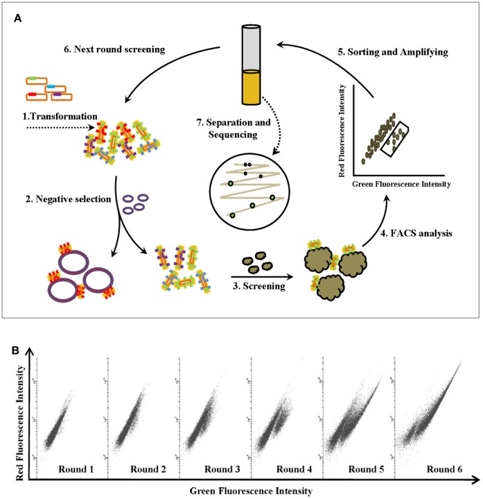
Currently in clinic, people use hematoxylin and eosin stain (H&E stain) and immunohistochemistry methods to identify the generation and genre of cancers for human pathological samples. Since these methods are inaccurate and time consuming, developing a rapid and accurate method to detect cancer is urgently demanded. In our study, binding peptides for lung cancer cell line A549 were identified using bacteria surface display method. With those binding peptides for A549 cells on the surface, the fluorescent bacteria (Escherichia coli with stably expressed green fluorescent protein) were served as specific detecting reagents for the diagnosis of cancers. The binding activity of peptide-fluorescent bacteria complex was confirmed by detached cancer cells, attached cancer cells and mice tumor xenograft samples. A unique fixation method was developed for peptide-bacteria complex in order to make this complex more feasible for the clinic use. This peptide-fluorescent bacteria complex has great potential to become a new diagnostic tool for clinical application.
Conflict of interest statement
Figures


Similar articles
-
Accurate and semi-automated analysis of bacterial association with mammalian cells.J Microbiol Methods. 2016 Mar;122:8-12. doi: 10.1016/j.mimet.2015.12.016. Epub 2016 Jan 6. J Microbiol Methods. 2016. PMID: 26769557
-
Peptide discovery using bacterial display and flow cytometry.Methods Enzymol. 2012;503:75-97. doi: 10.1016/B978-0-12-396962-0.00004-5. Methods Enzymol. 2012. PMID: 22230566
-
In vivo imaging of mesenchymal-epithelial transition factor (c-Met) expression using an optical imaging system.Bioconjug Chem. 2009 Jul;20(7):1299-306. doi: 10.1021/bc8005539. Bioconjug Chem. 2009. PMID: 19534520
-
Fluorescent proteins as visible in vivo sensors.Prog Mol Biol Transl Sci. 2013;113:389-402. doi: 10.1016/B978-0-12-386932-6.00010-7. Prog Mol Biol Transl Sci. 2013. PMID: 23244796 Review.
-
Flow cytometry of fluorescent proteins.Methods. 2012 Jul;57(3):318-30. doi: 10.1016/j.ymeth.2012.01.003. Epub 2012 Jan 24. Methods. 2012. PMID: 22293036 Review.
Cited by
-
Cytokine-induced killer cells co-cultured with non-cell derived targeting peptide-loaded dendritic cells induce a specific antitumor response.Cancer Biol Ther. 2019;20(5):720-728. doi: 10.1080/15384047.2018.1564561. Epub 2019 Feb 19. Cancer Biol Ther. 2019. PMID: 30777479 Free PMC article.
-
Reforming the Chimeric Antigen Receptor by Peptide Towards Optimized CAR T Cells With Enhanced Anti-Cancer Potency and Safety.Front Bioeng Biotechnol. 2022 Jun 17;10:928169. doi: 10.3389/fbioe.2022.928169. eCollection 2022. Front Bioeng Biotechnol. 2022. PMID: 35782491 Free PMC article.
-
Tumor-targeting peptides from combinatorial libraries.Adv Drug Deliv Rev. 2017 Feb;110-111:13-37. doi: 10.1016/j.addr.2016.05.009. Epub 2016 May 19. Adv Drug Deliv Rev. 2017. PMID: 27210583 Free PMC article. Review.
References
-
- Gao XH, Cui YY, Levenson RM, Chung LWK, Nie SM (2004) In vivo cancer targeting and imaging with semiconductor quantum dots. Nature Biotechnology 22: 969–976. - PubMed
-
- Wang F, Banerjee D, Liu YS, Chen XY, Liu XG (2010) Upconversion nanoparticles in biological labeling, imaging, and therapy. Analyst 135: 1839–1854. - PubMed
-
- Xu RX, Povoski SP, Martin EW (2010) Targeted delivery of microbubbles and nanobubbles for image-guided thermal ablation therapy of tumors. Expert Review of Medical Devices 7: 303–306. - PubMed
-
- Scott AM, Wolchok JD, Old LJ (2012) Antibody therapy of cancer. Nature Reviews Cancer 12: 278–287. - PubMed
-
- Graham MM, Menda Y (2011) Radiopeptide Imaging and Therapy in the United States. Journal of Nuclear Medicine 52: 56s–63s. - PubMed
Publication types
MeSH terms
Substances
LinkOut - more resources
Full Text Sources
Other Literature Sources
