. 2013;8(1):e54485.
doi: 10.1371/journal.pone.0054485. Epub 2013 Jan 22.

Identification of two evolutionarily conserved 5' cis-elements involved in regulating spatiotemporal expression of Nolz-1 during mouse embryogenesis

Affiliations

Identification of two evolutionarily conserved 5' cis-elements involved in regulating spatiotemporal expression of Nolz-1 during mouse embryogenesis

Sunny Li-Yun Chang et al. PLoS One. 2013.

Abstract

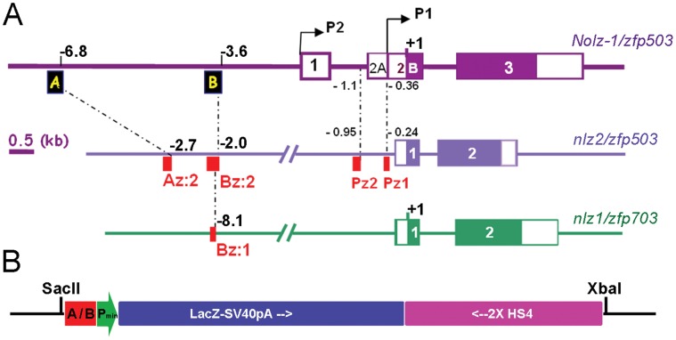
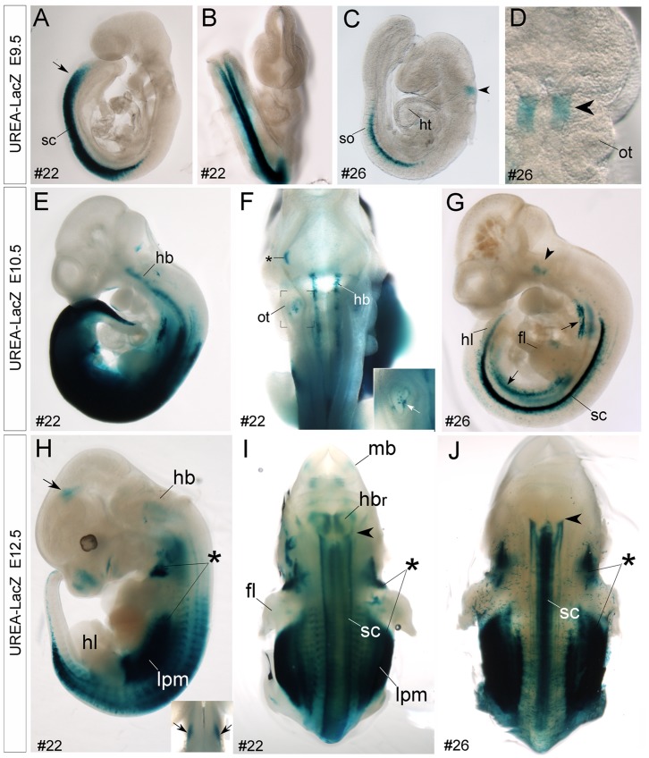
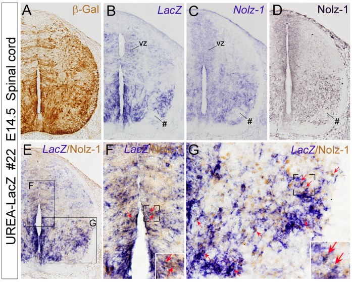
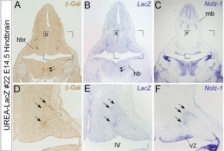
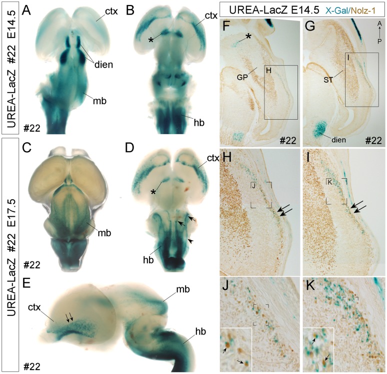
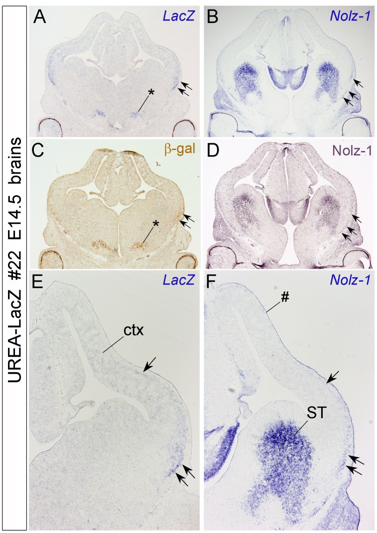
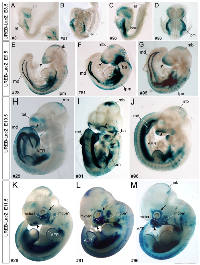
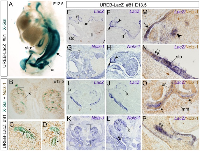
Proper development of vertebrate embryos depends not only on the crucial funtions of key evolutionarily conserved transcriptional regulators, but also on the precisely spatiotemporal expression of these transcriptional regulators. The mouse Nolz-1/Znf503/Zfp503 gene is a mammalian member of the conserved zinc-finger containing NET family. The expression pattern of Nolz-1 in mouse embryos is highly correlated with that of its homologues in different species. To study the spatiotemporal regulation of Nolz-1, we first identified two evolutionarily conserved cis-elements, UREA and UREB, in 5' upstream regions of mouse Nolz-1 locus. We then generated UREA-LacZ and UREB-LacZ transgenic reporter mice to characterize the putative enhancer activity of UREA and UREB. The results indicated that both UREA and UREB contained tissue-specific enhancer activity for directing LacZ expression in selective tissue organs during mouse embryogensis. UREA directed LacZ expression preferentially in selective regions of developing central nervous system, including the forebrain, hindbrain and spinal cord, whereas UREB directed LacZ expression mainly in other developing tissue organs such as the Nolz-1 expressing branchial arches and its derivatives, the apical ectodermal ridge of limb buds and the urogenital tissues. Both UREA and UREB directed strong LacZ expression in the lateral plate mesoderm where endogenous Nolz-1 was also expressed. Despite that the LacZ expression pattern did not full recapitulated the endogenous Nolz-1 expression and some mismatched expression patterns were observed, co-expression of LacZ and Nolz-1 did occur in many cells of selective tissue organs, such as in the ventrolateral cortex and ventral spinal cord of UREA-LacZ embryos, and the urogenital tubes of UREB-LacZ embryos. Taken together, our study suggests that UREA and UREB may function as evolutionarily conserved cis-regulatory elements that coordinate with other cis-elements to regulate spatiotemporal expression of Nolz-1 in different tissue organs during mouse embryogenesis.


Conflict of interest statement

Competing Interests: The authors have declared that no competing interests exist.

Figures

Figure 1
Figure 1. Evolutionarily conserved up-stream cis-elements of mouse Nolz-1/Zfp503 gene and the UREA-LacZ and UREB-LacZ transgenic constructs.
A: Diagrams show the locations of the UREA (A) and UREB (B) (black boxes) conserved upstream cis-elements of mouse Nolz-1/zfp503 and their relative conserved regions in zebrafish nlz2/zfp503 (red boxes, Az:2, Bz:2) and nlz1/zfp703 loci (red box, Bz:1). Pz1 and Pz2 (red boxes) are another two zebrafish/mouse conserved cis-elements identified in the potential promoter region of nlz2/zfp503. Numbers indicate the distances (in kb) relative to the translation start condon (+1). B: The UREA-LacZ and UREB-LacZ transgenic constructs. The UREA or UREB (A/B) elements were inserted upstream to a minimal promoter (Pmin) following by LacZ expression cassette (LacZ-SV40pA) and two direct repeats of insulators (2XHS4). The SacII and XbaI sites were used to release the transgenic fragments from the vector.
Figure 2
Figure 2. Detection of X-gal-positive signals in spinal cord and hindbrain of E9.5-E12.5 UERA–LacZ embryos.
A–J: Whole mount X-gal staining of E9.5 (A–D), E10.5 (E–G) and E12.5 (H–J) UREA-LacZ embryos. Prominent X-gal-positive signals are detected in the spinal cord (sc) of either line #22 (A, B, E, F) or #26 (C, D, G) UREA-LacZ embryos at E9.5 and E10.5. Note that, this expression is earlier and stronger in line #22 than in line #26. Interestingly, in the hindbrain (hb) region, X-gal-positive signals are detected in a selective domain of hindbrain (arrowhead) in line #26 embryos at E9.5 (C, D) and E10.5 (G). Later at E12.5, the X-gal-positive signals expend into more caudal regions of hindbrain with a pattern of longitudinal columns (arrowhead in J indicates the anterior expression border). In line #22 hindbrain, longitudinal X-gal-positive signals are detected from E10.5 at more broad regions than line #26 (E, F). H, I: By E12.5, the X-gal-positive signals are also detected in the rostral part of hindbrain (hbr, H). Asterisks in H-J indicate strong X-gal-positive signals in the paraxial mesenchyme of embryos in both lines. Notably, X-gal-positive signals are also present in the lateral plate mesoderm (lpm, H, I; arrows in G) in both lines, and in otic vesicle (ot, F and white arrow in inset at high magnification) and midbrain region (mb, I; arrows in H and inset of H) of line #22 embryos. Asterisk in F indicates a non-specific signal. fl, forelimb bud; ht, heart; hl, hindlimb bud; so, somites.
Figure 3
Figure 3. Detection of LacZ and Nolz-1 co-expressing cells in spinal cord of UREA-LacZ embryos.
A–D: Adjacent transverse sections of E14.5 spinal cord of line #22 UREA-LacZ embryos at the lumbar level. Expression patterns of β-gal protein (A) and LacZ mRNA (B) are similarly except that, in addition to soma, β-gal immunoreactivity is present in neurites, which results in broad expression pattern (A). Expression of Nolz-1 at mRNA (C) and protein (D) levels are consistently detected in selective domains of spinal cord, including in the ventricular zone (vz) of dorsal and ventral spinal cord and medial and lateral regions of the ventral spinal cord where LacZ gene (B) is also expressed. A LacZ and Nolz-1-negative domain is present in the ventrolateral part of spinal cord (see # in B–D). E–G: Double labeling of LacZ mRNA and Nolz-1 protein identifies many co-expressing cells (red arrows) in the ventricular zone (F) and in the medial and lateral domains of basal plate of spinal cord (G ). The boxed regions in E are shown at high magnification in F and G.
Figure 4
Figure 4. Detection of LacZ expression in a Nolz-1 expressing domain in rostral hindbrain of line #22 UREA-LacZ embryos.
A–C: Expressions of β-gal protein (A), LacZ mRNA (B), and Nolz-1 mRNA (C) on coronal hindbrain sections of line#22 E14.5 UREA-LacZ embryo. D–E: High magnification views of the bracketed regions in A–C, respectively. Expressions of LacZ mRNA and β-gal protein are both detected in a differentiated domain of rostral hindbrain (triple arrows in D, E) where endogenous Nolz-1 mRNA is expressed (F). LacZ expression is, however, absent in the nearby Nolz-1-expressing ventricular zone (vz, F). Double arrows in A and B indicate a domain in which β-gal expression is detected with little LacZ mRNA expression. IV, fourth ventricle; mb, midbrain.
Figure 5
Figure 5. Detection of X-gal-positive signals in specific brain regions and LacZ+/Nolz-1+ cells in the ventral cortex of line #22 UERA-LacZ embryos.
A–E: Whole mount X-gal staining of E14.5 (A, B) and E17.5 (C–E) embryonic brains. X-gal-positive signals are detected in the cerebral cortex (ctx, B, D), ventral forebrain (asterisk, B, D), diencephalon (dien, A), midbrain (mb, A, C, E) and hindbrain (hb, B, D, E) regions. D: Three X-gal-positive longitudinal columns (arrowhead, arrows) are found in the hindbrain. The arrows indicate the X-gal-positive columns which are also observed in the hindbrain of line #26 (Fig. S6), whereas the arrowhead indicates an additional X-gal-positive column found only in line #22. E: The double arrows indicate the ventral-to-dorsal gradient of X-gal-positive signals in the ventral cortex. A, C: dorsal view; B, D: ventral view; E: lateral view. F–K: Double labeling of X-gal-positive signal and Nolz-1 protein on E14.5 horizontal forebrain sections of line#22 UREA-LacZ embryos. Nolz-1 is expressed in the striatum (ST), but not in the globus pallidus (GP), nor in the X-gal-positive domains in ventral forebrain (asterisk, F) and diencephalon (dien, G). In the lateral cortex, a stream of X-gal-positive cells is observed. Within this cell stream, many X-gal-positive cortical cells co-expressing Nolz-1 (small arrows in insets of J, K) are detected. Anterior is to the top of figures F–K. Figure F is at ventral level and Figure G is at more dorsal level. The boxed regions in F, G and the bracketed regions in H, I, J, K are shown at high magnification in H, I, J, K, and insets in J, K, respectively. IV, fourth ventricle; mb, midbrain.
Figure 6
Figure 6. Detection of LacZ and Nolz-1 mRNA expression in the ventrolateral cortex of E14.5 line #22 UREA-LacZ brain.
A–D: Expression of LacZ mRNA (A), Nolz-1 mRNA (B), β-gal (C) and Nolz-1 protein (D) in coronal forebrain sections. Expressions of LacZ mRNA and β-gal protein are detected in a domain of ventral forebrain (asterisk, C) and in a strip of lateral cortex (ctx, double arrows in A, C and arrows in E). Nolz-1 mRNA is also expressed in a similar lateral strip in cortex (arrows in B, D, F). Note that, more LacZ- and Nolz-1-expressing cells are present in the ventral part (double arrows, E, F) than in the dorsal part (single arrow, F) of the strip. ST, striatum. # indicates non-specific staining of meninges.
Figure 7
Figure 7. Detection of X-gal-positive signals in specific tissue organs of early developing UERB-LacZ embryos.
A–D: X-gal signals are detected in the lateral plate mesoderm (lpm) and head fold structure (hf) of line #81 (A, B) and #96 (C, D) UREB-LacZ embryos at E8.5. E–G: At E9.5, the common regions containing X-gal-positive signals in all three lines of #28 (E), #81 (F) and #96 (G) are the anterior ridge of head (arrowhead), midbrain (mb), lateral plate mesoderm (lpm), mesonephric duct (md) and epidermis of embryonic bodies. Strong X-gal-positive signals are also detected in the branchial arches (ba) of line #81. H–M: Later at E10.5 (H–J) and E11.5 (K–M), prominent X-gal-positive signals are detected in the apical ectodermal ridge (AER, H, K, L) of limb buds and in olfactory pit (arrowhead, H–M). Although the expression levels are varied, selectively expression of LacZ in the branchial arches (ba) and its derivatives, including the mandibular (mdba1) and maxillary (mxba1) components of the first branchial arch, are consistently found in all three lines, (H–M), so as the selective expression of LacZ in the surrounding placodal tissues (arrow in H–M). A, C, E–H, J–M: side view; B, D, I: front view.
Figure 8
Figure 8. Detection of X-gal-positive signals in Nolz-1-expressing urogenital organs of UREB-LacZ embryos.
A: By whole-mount staining, X-gal-positive signals are detected in the urogenital duct (arrow), ureteric tubules (ur) and stomach (sto) of E12.5 line #81 embryos. B–C: Many X-gal+/Nolz-1+ cells (arrows in C, D) were detected in some renal tubules of E13.5 kidney (k). The boxed regions in B are shown at high magnification in C, D. E–L: Expressions of LacZ (E, F, I, J) and Nolz-1 mRNAs (G, H, K, L) in transverse sections of urogenital organs of E13.5 line #81 embryos. The bracketed regions in E, G, I, K are shown at high magnification in F, H, J, L, respectively. M–P: Double labeling of LacZ mRNA (purple) and Nolz-1 protein (brown). LacZ and Nolz-1 mRNAs are both detected in the genital ridge (arrow) and genital duct (arrowhead) (see also arrow and arrowhead in F, H). Many LacZ+/Nolz-1+ cells are detected in these genital tissues (M). LacZ mRNA is also detected in the adrenal gland (ad, E) and stomach (sto, E) of UREB-LacZ embryo. LacZ and Nolz-1 co-expressing cells in stomach are detected (arrows, N). In the embryonic kidney (k), LacZ mRNA is mainly expressed in tubular cells (I, J, O), whereas Nolz-1 mRNA (K, L) and protein are detected broadly both in the metanephric mesenchyme (mm, O) and in ureteric tubules (ur, O). LacZ and Nolz-1 co-expressing cells in ureteric tub are detected (arrows, O). Double arrows in L and P indicate Nolz-1 expression in urogenital tube where LacZ and Nolz-1 co-expressing cells are found. g, gonads; li, liver; ad, adrenal gland.

Similar articles

Cited by

References

    1. Nakamura M, Runko AP, Sagerstrom CG (2004) A novel subfamily of zinc finger genes involved in embryonic development. J Cell Biochem 93: 887–895. - PubMed
    1. Holland DG, Burleigh A, Git A, Goldgraben MA, Perez-Mancera PA, et al. (2011) ZNF703 is a common Luminal B breast cancer oncogene that differentially regulates luminal and basal progenitors in human mammary epithelium. EMBO Mol Med 3: 167–180. - PMC - PubMed
    1. Slorach EM, Chou J, Werb Z (2011) Zeppo1 is a novel metastasis promoter represses E-cadherin expression and regulated p120-catenin isoform expression and location. Genes Dev 25: 471–484. - PMC - PubMed
    1. Zhao X, Yang Y, Fitch DH, Herman MA (2002) TLP-1 is an asymmetric cell fate determinant that responds to Wnt signals and controls male tail tip morphogenesis in C. elegans . Development 129: 1497–1508. - PubMed
    1. Weihe U, Dorfman R, Wernet MF, Cohen SM, Milan M (2004) Proximodistal subdivision of Drosophila legs and wings: the elbow-no ocelli gene complex. Development 131: 767–774. - PubMed

Publication types

MeSH terms

LinkOut - more resources